分子生物学Chapter 2Protein Structure.docx
《分子生物学Chapter 2Protein Structure.docx》由会员分享,可在线阅读,更多相关《分子生物学Chapter 2Protein Structure.docx(16页珍藏版)》请在冰豆网上搜索。

分子生物学Chapter2ProteinStructure
Chapter2ProteinStructure
Introduction
Sizesandshapesofproteins
Globlarproteins,includingmostenzymes,behaveinsolutionlikecompact,roughlysphericalparticles.Fibrousproteinshaveahighaxialratioandareoftenofstructuralimportance,forexamplefibroinandkeratin.SizesrangefromafewthousandtoseveralmillionDaltons.
Someproteinscontainboundnonproteinmaterials(prostheticgroupsorothermacromolecules),whichaccountsfortheincreasedsizesandfunctionalitiesoftheproteincomplexs.
Shapesofproteins
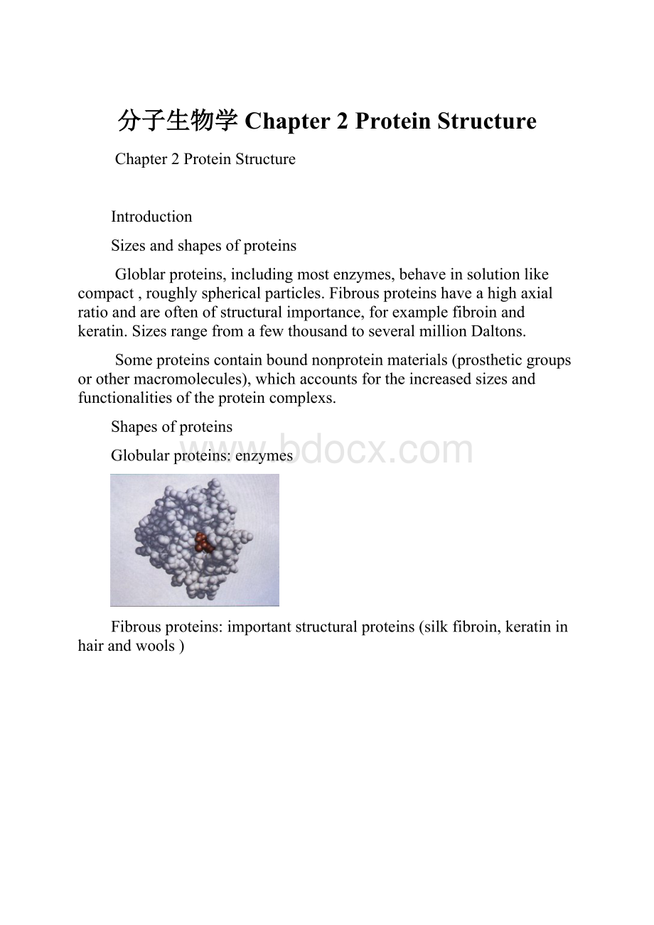
Globularproteins:
enzymes
Fibrousproteins:
importantstructuralproteins(silkfibroin,keratininhairandwools)
Chapter2ProteinStructure
1.AminpAcids
R-CH(NH2)-COOH
va-carbonischiral(asymmetric)exceptinglycine(RisH)
vAminoacidscanexitinbothD-andL-stereoisomers,butonlyL-isomersarefoundinproteins
vAminoacidsaredipolarions(zwitterions)inaqueoussolutionandareamphoteric.
vThesidechains(R)differinsize,shape,chargeandchemicalreactivity
vAfewproteinscontainnonstandardaminoacidsthatareformedbypost-translationalmodificationoftheparentaminoacids.
1.1Aminoacidswithchargedsidechains
“Basic”aminoacids:
containingpositivelychargedgroups
Lysine(Lys,K):
asecondaminogroupattachedtothee-carbonatom
Arginine(arg,R):
aguanidinogroupattachedtothed-carbonatom
Histidine(His,H):
aimidazolegrouphasapKanearneutrality.Thisgroupcanbereversiblyprotonatedunderphysiologicalconditions,whichcontributetothecatalyticmechanismofmanyenzymes.
1.2.Aminoacidswithpolarunchargedsidechains(hydrophilic)--containinggroupsthatformhydrogenbondswithwater
Serine(Ser,S)&threonine(Thr,T)havehydroxylgroups.
1.3.Aminoacidswithnonpolaraliphaticsidechains(hydrophobic)
Alkylsidechains
1.4.Aminoacidswitharomaticsidechains(hydrophobic)
2.Primarystructure
Aminoacidsarelinkedbypeptidebondsbetweenalpha-carboxylandalpha-aminogroups.TheresultingpolypeptidesequencehasanNterminusandaCterminus.
Polypeptidescommonlyhavebetween100and1500aminoacidslinkedinthisway
Formationofapeptidebond(shadedingray)inadipeptide.
vTheaminoacidinapeptideisalsocalledaresidue.
Thesmallestaminoacid,glycine,hasasinglehydrogenatomasitsRgroup.Itssmallsizeallowsittofitintotightspaces.Unlikeanyoftheothercommonaminoacids,prolinehasacyclicringthatisproducedbyformationofacovalentbondbetweenitsRgroupandtheaminogrouponCα.Prolineisveryrigid,anditspresencecreatesafixedkinkinaproteinchain.Prolineandglycinearesometimesfoundatpointsonaprotein'ssurfacewherethechainloopsbackintotheprotein
3.SecondaryStructure
vSecondarystructurereferstothelocalizedorganizationofpartsofapolypeptidechain,whichcanassumeseveraldifferentspatialarrangements.Asinglepolypeptidemayexhibitalltypesofsecondarystructure.Withoutanystabilizinginteractions,apolypeptideassumesarandom-coilstructure.
3.1Modeloftheahelix.
Thepolypeptidebackboneisfoldedintoaspiralthatisheldinplacebyhydrogenbonds(blackdots)betweenbackboneoxygenatomsandhydrogenatoms.Allthehydrogenbondshavethesamepolarity.Theoutersurfaceofthehelixiscoveredbytheside-chainRgroups.
a-helix
every3.6residuesmakeoneturn,
thedistancebetweentwoturnsis0.54nm,
theC=O(orN-H)ofoneturnishydrogenbondedtoN-H(orC=O)oftheneighboring
3.2BetaStrandandBetaSheet
vInaβstrand,thetorsionangleofN-Cβ-C-Ninthebackboneisabout120degrees. Thefollowingfigureshowstheconformationofanidealβstrand. Notethatthesidechainsoftwoneighboringresiduesprojectintheoppositedirectionfromthebackbone.
Betasheet
vAβsheetconsistsoftwoormorehydrogenbondedβstrands. Thetwoneighboringβstrandsmaybeparalleliftheyarealignedinthesamedirectionfromoneterminus(NorC)totheother,oranti-paralleliftheyarealignedintheoppositedirection.
bSHEETS.(a)Asimpletwo-strandedbsheetwithantiparallelbstrands.Asheetisstabilizedbyhydrogenbonds(blackdots)betweenthebstrands.Theplanarityofthepeptidebondforcesabsheettobepleated;hence,thisstructureisalsocalledabpleatedsheet,orsimplyapleatedsheet.(b)SideviewofabsheetshowinghowtheRgroupsprotrudeaboveandbelowtheplaneofthesheet.(c)ModelofbindingsiteinclassIMHC(majorhistocompatibilitycomplex)molecules,whichareinvolvedingraftrejection.Asheetcomprisingeightantiparallelbstrands(green)formsthebottomofthebindingcleft,whichislinedbyapairofahelices(blue).Adisulfidebondisshownastwoconnectedyellowspheres.TheMHCbindingcleftislargeenoughtobindapeptide810residueslong.
3.3Turns
Composedofthreeorfourresidues,turnsarecompact,U-shapedsecondarystructuresstabilizedbyahydrogenbondbetweentheirendresidues.Theyarelocatedonthesurfaceofaprotein,formingasharpbendthatredirectsthepolypeptidebackbonebacktowardtheinterior.Glycineandprolinearecommonlypresentinturns.
4.TertiaryStructure
vTertiarystructure,thenext-higherlevelofstructure,referstotheoverallconformationofapolypeptidechain,thatis,thethree-dimensionalarrangementofalltheaminoacidsresidues.Incontrasttosecondarystructure,whichisstabilizedbyhydrogenbonds,tertiarystructureisstabilizedbyhydrophobicinteractionsbetweenthenonpolarsidechainsand,insomeproteins,bydisulfidebonds.Thesestabilizingforcesholdtheαhelices,βstrands,turns,andrandomcoilsinacompactinternalscaffold
Theribbonrepresentationofthe3DstructureofRNaseA.
vNoncovalentinteractionbetweensidechainsthatholdthetertiarystructuretogether:
vanderWaalsforces,hydrogenbonds,electrostaticsaltbridges,hydrophobicinteractions
vCovalentinteraction:
disulfidebonds
vDenaturationofproteinbydisruptionofits2oand3ostructurewillleadtoarandomcoilconformation
5.Quaternarystructure
Manyproteinsarecomposedoftwoormorepolypeptidechains(subunits).Thesesubunitsmaybeidenticalordifferent.Thesameforceswhichstabilizetertiarystructureholdthesesubunitstogether.Thisleveloforganizationcalledquaternarystructure.
Prostheticgroups
vProstheticgroupscovalentlyornoncovalentlyattachedtomanyconjugatedproteins,andgivetheproteinschemicalfunctionality.Manyareco-factorsinenzymereactions.
vExamples:
hemegroupsinhemogobin
Organizationofthecataboliteactivator
protein(CAP)
6.Domains,motifsandfamilies
6.1ProteinMotifs
vAmotifisacharacteristicdomainstructureconsistingoftwoormoreαhelicesorβstrands.
vCommonexamplesincludecoiledcoil,helix-turn-helixhelix-loop-helix,zincfinger,leucinezipperetc.
vManyproteinscontainoneormoremotifsbuiltfromparticularcombinationsofsecondarystructures.
vAmotifisdefinedbyaspecificcombinationofsecondarystructuresthathasaparticulartopologyandisorganizedintoacharacteristicthree-dimensionalstructure.
Structuralmotifs:
•Groupingsofsecondarystructuralelementsthatfrequentlyoccuringlobularproteins
•Oftenhavefunctionalsignificanceandrepresenttheessentialpartsofbindingorcatalyticsitesconservedamongaproteinfamily
•Representthebestsolutiontoastructural-functionalrequirement
βαβmotif
6.2Domains:
Structurallyindependentunitsofmanyproteins,connectedbysectionswithlimitedhigherorderstructurewithinthesamepolypeptide.
Theycanalsohavespecificfunctionsuchassubstratebinding
FunctionalDomains
vDomainssometimesaredefinedinfunctionaltermsbasedonobservationsthattheactivityofaproteinislocalizedtoasmallregionalongitslength.Forinstance,aparticularregionorregionsofaproteinmayberesponsibleforitscatalyticactivity(e.g.,akinasedomain)orbindingability(e.g.,aDNA-bindingdomain,membrane-bindingdomain).
Modules:
vTheorganizationoftertiarystructureintodomainsfurtherillustratestheprinciplethatcomplexmoleculesarebuiltfromsimplercomponents.Likesecondary-structuremotifs,tertiary-structuredomainsareincorporatedasmodulesintodifferentproteins,therebymodifyingtheirfunctionalactivities.Themodularapproachtoproteinarchitectureisparticularlyeasytorecognizeinlargeproteins,whichtendtobeamosaicofdifferentdomainsandthuscanperformdifferentfunctionssimultaneously
Schematicdiagramsofvariousproteins,illustratingtheirmodularnature.
vEpidermalgrowthfactor(EGF)isgeneratedbyproteolyticcleavageofaprecursorproteincontainingmultipleEGFdomains(orange).TheEGFdomainalsooccursinNeuproteinandintissueplasminogenactivator(TPA).Otherdomains,ormodules,intheseproteinsincludeachymotrypticdomain(purple),animmunoglobulindomain(green),afibronectindomain(yellow),amembrane-spanningdomain(pink),andakringledomain(blue).
6.3Proteinfamilies:
Structurallyandfunctionallyrelatedproteinsfromdifferentsources
Evolutionarytreeshowinghowtheglobinproteinfamilyarose,startingfromthemostprimitiveoxygen-bindingproteins,leghemoglobins,inplants.Sequencecomparisonshaverevealedthatevolutionoftheglobinproteinsparallelstheevolutionofvertebrates.Majorjunctionsoccurredwiththedivergenceofmyoglobinfromhemoglobinandthelaterdivergenceofhemoglobinintotheaandbsubunits.
Domains,motif