实验八聚丙烯酰胺凝胶圆盘电泳.docx
《实验八聚丙烯酰胺凝胶圆盘电泳.docx》由会员分享,可在线阅读,更多相关《实验八聚丙烯酰胺凝胶圆盘电泳.docx(12页珍藏版)》请在冰豆网上搜索。
实验八聚丙烯酰胺凝胶圆盘电泳
实验八聚丙烯酰胺凝胶圆盘电泳
【实验目的】
1.掌握盘状聚丙烯酰胺凝胶电泳的基本原理。
2.学习盘状聚丙烯酰胺凝胶电泳的操作技术,用于分离蛋白质。
【实验原理】
CH2=CH
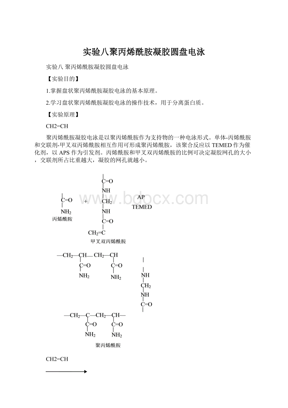
聚丙烯酰胺凝胶电泳是以聚丙烯酰胺作为支持物的一种电泳形式。
单体-丙烯酰胺和交联剂-甲叉双丙烯酰胺相互作用可形成聚丙烯酰胺,该聚合反应以TEMED作为催化剂,以APS作为引发剂。
丙烯酰胺和甲叉双丙烯酰胺的比例可决定凝胶网孔的大小,交联剂所占比重越大,凝胶的网孔就越小。
CH2=CH
—CH2—CH—
CH2—CH—
利用聚丙烯酰胺凝胶电泳进行蛋白质分离主要依据以下两个因素:
蛋白质所带静电荷:
在不同的pH条件下蛋白质所带电荷不同。
在一定的电场条件下蛋白质将向与其所带电荷相反的电极方向移动,移动速率取决于蛋白质表面电荷的数量,电压越强或电荷越多则蛋白质移动的越远。
其次,蛋白质的形状和大小:
蛋白质在电泳中所受的阻力主要取决于样品的大小与凝胶网孔大小之间的关系。
蛋白质分子越小或凝胶网孔越大,所分离样品所受阻力就愈小,则在电场中的迁移率就越大。
在非变性电泳中,天然蛋白质的分离就是蛋白质所带电荷、分子大小及分子形状等因素共同影响,作用的结果。
电压x样品静电荷
迁移率=
摩擦力
浓缩效应可显著提高聚丙烯酰胺凝胶电泳的分辨率,该效应可通过引入浓缩胶和不连续缓冲溶液系统而获得。
浓缩胶处于分离胶的顶部,因此样品在进入分离胶之前首先要经过浓缩胶。
它是由较低浓度的丙烯酰胺构成,当样品经过浓缩胶时由于胶内网孔较分离胶网孔大,样品的移动速度较快,最终使样品“堆积”在浓缩胶和分离胶之间。
另外,浓缩胶还具有比分离胶更低的pH。
浓缩胶内的缓冲溶液是Tris-HCl(pH6.7),该pH远低于Tris的pK值(8.1)。
分离胶内的缓冲溶液是pH8.9的Tris-HCl,而电泳正负两极的缓冲溶液均为pH8.3的Tris-甘氨酸缓冲溶液。
在浓缩胶中,小分子并带有大量负电荷的Cl-在凝胶中的移动速率较快,而电荷较少且分子量较大的甘氨酸的移动速率较慢。
由于二者在同一电场中,具有相同的电流,由此形成的电压梯度导致甘氨酸离子始终跟随在Cl-的后面,带有负电荷的蛋白质样品则浓缩在甘氨酸离子和Cl-之间向正极移动。
当样品进入pH8.9的分离胶时,甘氨酸的解离度增加,在电场中的迁移率高于样品,蛋白质不再夹于两种离子之间向正极移动,这时的蛋白质依靠分子筛效应和电荷效应进行分离。
样品缓冲溶液
Tris/HClpH6.7
Tris/甘氨酸pH8.9
-
+
Tris/甘氨酸pH8.9
分离胶
Tris/HClpH8.9
浓缩胶
Tris/HClpH6.7
【实验材料】
1.实验器材
Bio-Rad公司Mini-ProteanⅡ型电泳仪;电源(电压200V,电流500mA);100℃沸水浴;Eppendorf管;微量注射器(50μl或100μl);带盖的玻璃或塑料小容器;摇床。
2.实验试剂
试剂号
配方
pH
1
分离胶缓冲溶液1mol/LHCl48.0ml
三羟甲基氨基甲烷36.3g
加蒸馏水到100ml
8.9
2
单体交联剂丙烯酰胺(Acr)30g
甲叉丙烯酰胺(Bis)0.8g
加蒸馏水到100ml
3
过硫酸铵100mg/ml
4
四甲基乙二胺(TEMED)
5
浓缩胶缓冲溶液1mol/LHCl48ml
三羟甲基氨基甲烷5.89g
加蒸馏水到100ml
6.7
6
电极缓冲溶液甘氨酸28.8g
三羟甲基氨基甲烷6.0g
加蒸馏水到1000ml,用前稀释10倍
8.3
7
染色液CBBG-2500.1g溶于95%乙醇后,加蒸馏水到100ml
8
示踪染料溴酚蓝0.05g溶于100ml蒸馏水
9
样品:
蛇毒干粉200mg溶于20ml,pH6.7缓冲溶液(5)中,再加入25%蔗糖20ml和溴酚蓝(8)10ml
凝胶溶液的配方
试剂
分离胶(ml)
浓缩胶(ml)
1
2
5
蒸馏水
3
4
1.25
2.5
-
6.195
0.05
0.005
-
1.0
1.25
7.64
0.10
0.005
总体积(ml)
10
10
Aa浓度(g%)
7.5
3.0
【实验操作】
1.凝胶柱的制备
取l0cm×0.6cm的玻璃管,选择较平整的一端为底端,量取7.5cm、8cm两处,画线;底端管口用小块胶布封口,插入橡皮垫中,垂直放置于试管架中。
用巴斯德滴管吸取分离胶,缓慢贴壁加胶到管内7.5cm处,立即加蒸馏水至8cm处。
待分离胶凝固后,将胶面上的水分甩掉,残留的水分用滤纸条吸干,用滴管速加浓缩胶到分离胶面上至8cm处,再小心地加一层覆盖水。
2.安装电泳槽
选择无气泡、无裂缝、长度合适的小玻璃管,撕掉胶布,安装到电泳仪上,注意要紧密,以防止上槽缓冲溶液漏液;向下槽注入电极缓冲液,注意用弯头滴管除去玻璃管下端的气泡;再向上槽倒入电极缓冲液淹没小管,同样用滴管除去气泡。
3.加样
用微量注射器取样品液30μl,沿壁加在浓缩胶面上,注入时要慢,避免激起电极缓冲液。
4.电泳
连接电极,上槽与负极相连,下槽与正极相连;调节电流为lmA/管,待示踪染料进入分离胶时调节电流为2mA/管,待示踪染料接近凝胶管底部约0.5cm处,切断电源,电泳时间为2小时~3小时。
5.剥胶
取下凝胶管,用局麻针头注射器吸取一定量的蒸馏水,将针头插入胶柱-与管壁之间,边注水边旋转玻管,直至胶柱与管壁分开,然后用洗耳球轻轻在玻管的一端加压,使凝胶柱从玻管缓慢滑出,将凝胶柱置于编号的试管内,用蒸馏水冲洗几次。
6.染色
将考马斯亮蓝染色剂倒入放有胶条的小试管中,没过胶条;60℃水浴保温40分钟~50分钟后取出,用水冲洗2次~3次,观察结果。
【实验结果】
观察凝胶条中的蛋白质与考马斯亮蓝染色剂结合后所形成的蓝色复合物,并通过画图记录结果。
【思考题】
⒈聚丙烯酰胺凝胶电泳分离生物大分子的基本原理?
⒉样品液中加入蔗糖和溴酚蓝的目的是什么?
Experiment8PolyacrylamideGelElectrophoresisinCylindricalTube
【Purpose】
1Mastertheprinciplepolyacrylamidegelelectrophoresis.
2Learntousethisapproachoftube-polyacrylamidegelelectrophoresistoseparatethetoxinofsnake.
【Principle】
CH2=CH
Polyacrylamidegelelectrophoresisisakindofelectrophoresisinwhichthesupportmediaispolyacrylamide(PAGE).Theorganicmonomer,acrylamidecanreactwithcross-linkingreagent,methylenebisacrylamidetoformpolyacrylamidegel.ThispolymerizingreactionneedsTEMEDascatalystandAPSasarisingagent.
CH2—CH—
—CH2—CH—
CH2=CH
Theproportionbetweenacrylamideandmethylenebisacrylamideresultsinthesizeofporesinthegel,smallerporessizesareobtainedbyusingahigherconcentrationofcross-linkingreagenttoformthegel.
Theseparationofproteininpolyacrylamidegelelectrophoresisdependsontwoaspects:
Netchargesontheproteins:
DependingonthepHofthebuffer,proteinsinasamplewillcarrydifferentcharges.Whenanelectricfieldisapplied,proteinswillmigratetowardstheircorrespondingpoles.Therateofmigrationwilldependonthestrengthoftheirnetsurfacecharges.Thehigherthevoltageorthegreaterthechargeontheproteinthefurtheritwillmove
ShapeandsizeoftheProteins:
Thefrictionofaproteinislargelydeterminedbytherelationshipbetweentheeffectivesizeofthemoleculeandthesizeoftheporesinthegel.Thesmallerthesizeofthemolecule,orthelargerthesizeoftheporesinthegel,thelowertheresistanceandthereforethefasteramoleculemovesthroughthegel.
Innon-denaturingelectrophoresis,thenativeproteinsareseparatedbasedonacombinationoftheircharge,sizeandshape.
appliedvoltagexmolecularcharge
mobility=
molecularfriction
Theresolutionofseparationinelectrophoresiscanbeimprovedbytheuseofastackinggelandadiscontinuousbuffersystem,whichcontributestothestackingeffect.
Thestackinggelresidesontopoftherunninggel,andthusthesamplepassesfirstthroughthestackinggel.Stackinggelismadeusingalowerpercentageofacrylamidethantherunninggelandithaslessmolecularsieving.So,afterloadingsamples,theproteinsrunrapidlythroughthestackinggelwhichishighlyporousandthen"stack"upattheinterfacebetweenthetwogelssincetherunninggelhasmuchsmallerpores.
Tris/GlycinepH8.9
ThestackinggelalsohasalowerpHthantherunninggel.ItisaTris-HClbufferatpH6.7,whichismuchbelowthepKofTris(8.1).TherunninggelisaTris-HClbufferatpH8.9andtherunningbufferforthegeloverallisTris-glycinepH8.3.Instackinggel,thefully-chargechloridewillmovefastthroughtheporous.Thelargerandslightlychargedglycinewillmoveslowly.Butthecurrentmustbethesamethroughoutthiselectricalcircuit.Avoltagegradientallowstheglycinetoremainjustbehindthechlorideions.Theproteinswithnegativechargewillmigratebetweenthechlorideandtheglycine,inverysharpbands.WhenthesamplesentertherunninggelwiththepHincreasingto8.9,theglycinebecomesmoresignificantlydeprotonatedanditmovesaheadoftheproteins.Theproteinsarenownotforcedtostackbetweenthetwoions,andcanproceedtobeseparatedbythemolecularsievingofthehigherconcentrationgel.
【Materials】
1.Apparatus
Bio-RadMini-Proteanapparatus;Powersupply(capacity200V,500MA);Boilingwaterbath;Eppendorfcentrifuge(optional);HamiltonSyringes(50μland100μlcapacity);Smallglassorplasticcontainerwithlid(i.e.12cm×16cm×3cm);Rockingorrotaryshaker.
2.Reagents
regent
menu
pH
1
Separatinggelbuffer:
1mol/LHCl48.0ml,Tris36.3g,adddistilledwaterto100ml.
8.9
2
Stockacrylamidesolution:
30gacrylamide,0.8gbis-acrylamide.Makeupto100mlindistilledwater.
3
10%Ammoniumpersulfateindistilledwater.
4
N,N,N',N'-tetramethylethylenediamine(TEMED).
5
Stackinggelbuffer:
48ml1mol/LHCl,Tris5.89gadddistilledwaterto100ml.
6.7
6
10×Electrophoresisbuffer:
Dissolve6.0gofTrisbaseand28.8gofglycineinwaterandadjustthevolumeto1L.
8.3
7
CoomassieGelStain:
Add0.1gCoomassieBlueR-250into45mlmethanol,45mlH20and10mlglacialaceticacid.
8
Footprintdyebromophenolblue:
bromophenolblue0.05gin100mldistilledwater.
9
Sample:
toxinofsnake200mgdissolvein20ml,pH6.7stackinggelbuffer,andadd25%sucrose20mlandbromophenolblue10ml.
PreparationofAcrylamideSolutions
regent
separatinggel(ml)
Stackinggel(ml)
1
2
5
Distilledwater
3
4
1.25
2.5
-
6.195
0.05
0.005
-
1.0
1.25
7.64
0.10
0.005
Totalvolumes(ml)
10
10
Theconcentrationofthegel(g%)
7.5
3.0
【Procedures】
1.Preparationofthegel
Cleantheinternalsurfacesoftheglasstube(10cm×0.6cm)anddry,thenmakemarkersat7.5cmand8.0cm.Envelopthebottomwithplastertightly,thenputitintocushion,andclampitinaverticalposition.UsingaPasteur(orlarger)pipettotransferseparatinggelmixturetothetubebyrunningthesolutioncarefullydowntheedge.Continuetoaddthissolutionuntilitreachestheposition7.5cm,gentlylaydistilledwaterontopoftheseparatinggeluntiltheposition8.0cmimmediately.Whenthepolymerizationhasfinishedinrunninggel,pourofftheoverlayingwaterandthenaddthestackinggelsolutiontothetubeuntilthesolutionreachestotheposition8.0cm.gentlylayabout0.5cmofdistilledwaterontopofthestackinggel.Thesamepointalsoshouldbenoticedaslaststepwhichpreventsdistilledwaterfrommixingwithgelsolution.Allowstackinggeltopolymerize(about30minutes).
2.Theinstallationofthegel
Placetheeligiblegeltubeintoelectrophoresischamber,verticallyandairtightly.Discardthebottomplaster,andmovethebubbleunderthebottom.Addelectrophoresisbuffertoinnerandouterreservoir,makingsurethatbothtopandbottomofgelareimmersedinbuffer.
3.LoadingSamples
Load30μlsamplesolutionwithminimsyringe,carefullydowntheedgetothesurfaceofstackinggel.
4.RunningaGel
Attachelectrodeplugstoproperelectrodes,Anodeshouldconnectwithinnerreserviorandcurrentshouldflowtowardstheanode;Turnonpowersupplyto1mA/tubewhenthedyemigratesinstackinggelandchangethecurrentto2mAwhenthedyemigratestoseparatinggel.Whenthedyemigratesto0.5cmfromthebottomofthegel,turnoffpowersupply.
5.Peeloffthegel
Removecylindricaltubefromelectrodeassemblyfirstlyanduseasyringewith10cmlongpinheadfullofwateraslube.Putthelongpinheadintothespacerbetweengelandglass,injectingandturninguntilthegelisseparatedfromthetube,andblowgentlytomakethegelslipout.
6.StainingtheGel
PutthegelintolittletubewithCoomassiebrilliantblue,andincubateat60℃for40min-50min,thentakeoutandwashwithwaterfortwoorthreetimes.
【Result】
ObservethebluecompoundwhichformedbetweenproteinandCoomassiebrilliantblueinthegelanddrawouttheresult.
【Questions】
⒈Whatistheseparatingprincipleofp
 升级会员
升级会员 