Mitral valve prolapse in the dog a model of mitral valve prolapse in manWord文件下载.docx
《Mitral valve prolapse in the dog a model of mitral valve prolapse in manWord文件下载.docx》由会员分享,可在线阅读,更多相关《Mitral valve prolapse in the dog a model of mitral valve prolapse in manWord文件下载.docx(33页珍藏版)》请在冰豆网上搜索。
,
Jens
Hä
ggströ
m
DOI:
http:
//dx.doi.org/10.1016/S0008-6363(00)00113-9
234-243
Firstpublishedonline:
1August2000
∙Article
∙Figures&
data
∙Information
∙Explore
∙PDF
Keywords
∙Aging
∙Epidemiology
∙Reninangiotensinsystem
∙Ultrasound
∙Valve(disease)
Timeforprimaryreview31days.
1Introduction
Mitralvalveprolapse(MVP),i.e.abnormalsystolicprotrusionofmitralvalveleafletsintotheleftatrium,isacommoncauseofseveremitralregurgitation(MR)requiringoperationinpeoplelivinginindustrializednations
[1,2].MVPhasbeenreportedtohavemanycausesbutinthemajorityofcasesitisaprimarycondition(calledprimaryMVPinthispaper)characterizedbyaprogressivemyxomatousdegenerationofthemitralvalveleafletsandchordaetendineae
[1–3].ThediseasetypicallyemergesinadolescencebutcomplicationssuchassevereMRusuallydonotoccuruntilmiddleageorsenescence
[1–3].Ananimalmodelwithashortercourseofdiseasecouldbeusefulinseveralways,forinstance,bymakingitfeasibletoevaluatetheeffectsofdifferentdrugsondiseaseprogression.Despitethis,noanimalmodelofprimaryMVPhasbeendescribedsofar.二尖瓣脱垂(MVP),即异常收缩凸出二尖瓣瓣叶进入左心房,是严重的二尖瓣关闭不全(MR),需要在生活在工业化国家[1,2]人操作的常见原因。
MVP已报道有很多原因,但在大多数情况下,它是一个基本条件(称为本文初级MVP)的特征在于以下二尖瓣瓣叶和腱索[1-3]渐进粘液变性。
这种疾病通常出现在青春期,但并发症,如严重的MR通常不会发生,直到middleage或衰老[1-3]。
与疾病的较短过程的动物模型可以在几个方面是有用的,例如,通过使其可行评估不同药物对疾病进展的影响。
尽管这样,初级的MVP无动物模型已经描述至今。
Frompathologicalstudies,ithaslongbeenknownthatmostdogsdevelopmyxomatousmitralvalvediseasewithageandthatthisdiseaseisverysimilarmacroscopicallyaswellasmicroscopicallytoprimaryMVPinhumans
[4,5].Traditionally,however,thecaninediseasehasbeengivennamesotherthanMVP,includingendocardiosisandchronicvalvulardisease.Recently,anumberofstudies,includingmanybasedonwell-definedechocardiographiccriteriaforthediagnosisofMVP,haveincreasedourunderstandingofthisdiseaseinthedog.Thepurposeofthisarticleistocomparetheknowledgewhichhasbeenaccumulatedaboutmyxomatousmitralvalvedisease/MVPinthedogwithknowledgeofprimaryMVPinhumans.从病理研究,人们早已知道,大多数狗开发粘液二尖瓣病变随着年龄和这种疾病非常相似宏观以及微观到在人类[4,5]主MVP。
传统上,然而,犬疾病已经给出的名称比MVP,包括心内膜炎和慢性瓣膜病等。
最近,一些研究,包括许多基于定义良好的超声心动图标准MVP的诊断,也增加了我们的这种疾病在狗的理解。
这篇文章的目的是比较已积累了约粘液二尖瓣病变/MVP与原发性MVP在人类知识的狗的知识。
2Pathology
Pathologically,primaryMVPinhumansisverysimilartocaninemyxomatousmitralvalvedisease
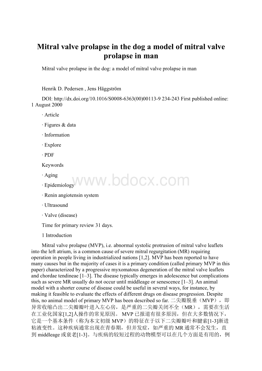
[4,5].Inbothspecies,theprincipalmacroscopicfindingsareenlarged,thickenedleaflets,interchordalhoodingandelongatedchordaetendineae(Fig.1A,B)
[4–9].Inaddition,affectedindividualsfrombothspeciesoftendisplaysimilar,albeitminor,changesinthetricuspidvalveand,inlatestages,secondaryfibrosisoftheleaflets,rupturedchordaetendineae,jetlesionsanddilationoftheleftventricle,leftatriumandmitralannulus
[4–9].Lightmicroscopyoftheleafletsshowsdepositionofglycosaminoglycansanddisruptionofcollagenastheprimaryfindingsinbothspecies(Fig.1C,D)
[5–7,9].Electronmicroscopyrevealsareaswithsparse,disorganizedcollagenfibrilswhichoftenhaveaspiralingappearance,indogsaswellasinhumans(Fig.1E,F)
[10].病理学上,在人类初级MVP的非常相似,犬粘液二尖瓣病变[4,5]。
在这两个物种中,主要宏观结果被放大,加厚传单,interchordal戴头罩和细长腱索(图1A,B)[4-9]。
此外,来自这两个物种受影响的个体经常显示类似,尽管轻微,在三尖瓣和变化,在后期,小叶的次级纤维化,破裂腱索,喷病变和左心室扩张,左心房和二尖瓣环[4-9]。
小叶的光学显微镜表明糖胺聚糖的沉积和胶原的破坏如在这两个物种的主要结果(图1C,D)[5-7,9]。
电子显微镜揭示了具有稀疏,紊乱的胶原纤维,往往有一个螺旋的外观,在狗中,以及在人类(图1E,F),[10]的区域。
∙Downloadfigure
∙Openinnewtab
∙Downloadpowerpoint
Fig.1
Myxomatousmitralvalvedisease/mitralvalveprolapseinthedog(A,CandE)andinman(B,DandF).Thephotographsonthetop(AandB)showpost-mortemspecimensfromadogandahuman,respectively.Themitralvalveleafletsareenlarged,thickenedanddisplayinterchordalhooding(arrows).Thejetlesionpresentontheatrialwallofthedog(arrowheads)resultfromimpactofregurgitantjetsofblood.Thephotographsinthecenter(CandD)showhistologysectionsoftheposteriormitralvalveleafletfromadogandahuman,respectively.ThesectionsarestainedwithPAS–Alcianblue–hematoxylin.Inbothvalves,severedepositionofglycosaminoglycans(blue)anddisrupted,disoganizedcollagen(pink)isseen.Scalebars:
1mm.Theelectronmicrographsonthebottom(EandF)arefromamyxomatousmitralvalveleafletfromadogandahuman,respectively.Collagenfibresarefragmentedandshowspiralingappearanceinlongitudinalsections(arrows).Cellulardebrisisseeninbetweenthecollagenbundles.Originalmagnification×
3500(E)and×
27 000(F).(B:
courtesyofUlrikBaandrup,MD,PhD;
C:
courtesyofToma
 升级会员
升级会员 