病理学诊断基础图片 1.docx
《病理学诊断基础图片 1.docx》由会员分享,可在线阅读,更多相关《病理学诊断基础图片 1.docx(12页珍藏版)》请在冰豆网上搜索。
病理学诊断基础图片1
病理学诊断基础图片
病理基本病变是病理诊断的基础,这里收集了一部分但愿对初学者有一定帮助。
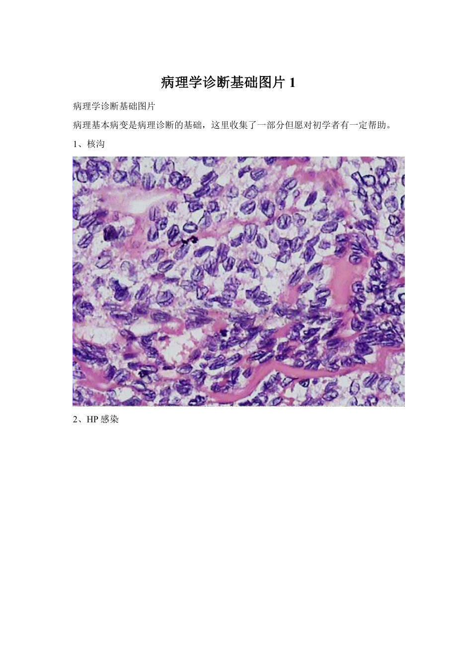
1、核沟
2、HP感染
3、腱鞘巨细胞瘤中的“多核巨细胞”及“含铁血黄素”
4、泡状核细胞(最醒目的核呈空泡状、核仁嗜酸的大细胞)
5、菊形团(五星指示)
6、泡沫细胞
7、肝细胞小泡型脂肪变
8、血栓
9、肝细胞核空泡变性(糖原核)
10、梭形细胞的栅栏状排列
11、正常鳞状上皮细胞之间的细胞间桥
12、玻璃样变性
13、子宫内膜腺癌
图示子宫内膜腺癌。
棕褐色不规则的肿块充满子宫腔并向肌层浸润。
大多数病人发病年龄在55-60岁之间,极少发生于40岁以下。
主要危险因素是长期过多雌激素的刺激。
雌激素升高导致内膜增生,并促使癌发生。
无排卵性月经周期、肥胖、分泌雌激素的卵巢肿瘤、未育、外源性雌激素治疗,都能增加子宫内膜腺癌发生的危险。
高血压、糖尿病也是其危险因素。
绝经期后不规则的阴道流血可能是其仅有的征象。
子宫无明显增大。
大多数子宫内膜腺癌局限于子宫(即Ⅰ期),其五年存活率为90%。
Theirregulartanmassfillingtheendometrialcavityandinfiltratingintothemyometriumoftheuterusisanendometrialadenocarcinoma,seentobemoderatelydifferentiatedmicroscopically.Mostpatientswiththisneoplasmare55to65yearsofage,andrarelyyoungerthan40years.Themajorriskfactorforthiscarcinomastemsfromprolongedestrogenstimulation.Conditionsthatleadtoincreasedestogenicexposurecanproduceendometrialhyperplasia,fromwhichacancercanarise.Anovulatorycycles,obesity,estrogen-producingovariantumors,lowparityornulliparity,andexogenousestrogentherapycanallincreasetheriskforendometrialadenocarcinomainthismanner.Hypertensionanddiabetesmellitusarealsoriskfactors.Irregularpostmenopausalbleedingmaybetheonlysign,andtheuterusmaynotbesignificantlyenlarged.Mostendometrialadenocarcinomasareconfinedtotheuterus(StageI)witha5-yearsurvivalaround90%.
14胆石症
伴有胆固醇结石的胆石症患者。
图示:
剖开的胆囊内可见大量混合石(胆石分为色素性胆石、胆固醇性胆石和混合石三种)。
大多数结石由钙、胆红素和胆固醇组成。
明显的浅黄色与主要成分胆固醇有关。
老龄化、女性、肥胖均为胆石症的危险因素。
伴结石形成的慢性胆囊炎往往有胆囊粘膜及胆囊壁的慢性炎症。
虽然大多数胆石症病人无临床表现,然而胆囊炎急性发作可致右上腹痛。
色素石主要由胆红素钙形成,主要见于溶血和胆道梗阻。
Agallbladderisopenedtorevealnumerouscompositegallstones.Moststonesaremixturesofcalcium,bilirubin,andcholesterol.Thisischolelithiasiswith"cholesterolstones."Theprominentyellowishcolortothesestonesisconsistentwiththeirpredominantcholesterolcomponent.Aging,femalesex,andobesityincreasetheriskforgallstoneformation.Therecanbechronicinflammationinthemucosaandwallofthegallbladderaccompanyingthestones--chroniccholecystitis.Thoughmostpersonswithgallstonesareasymptomatic,theremaybeepisodesofacutecholecystitismarkedbyintenserightupperquadrantpain.Theso-called"pigmentstones"composedmainlyofdarkcalciumbilirubinateareseeninpopulationswithchronichemolysis(hemoglobinopathies)andbiliarytractinfections.
15硬下疳梅毒螺旋体
性传播疾病梅毒患者病损硬下疳中可见梅毒螺旋体。
病原体为苍白密螺旋体。
晚期妊娠患者可传播给胎儿,从而使新生儿患先天性梅毒。
病变组织学特征为动脉内膜炎和浆细胞浸润性炎。
早期梅毒的重要特征是感染梅毒螺旋体两周后在原始部位形成硬下疳。
自愈后发生二期梅毒,持续两个月,其特征性病变是梅毒疹,随后进入静止期。
但经十几年的发展可出现三期梅毒的典型病损如神经系统梅毒、心血管梅毒或梅毒瘤。
此期很难查见病原体。
Seenherearespirochetesfromalesionofprimarysyphilisknownachancre.ThecausativeorganismisTreponemapallidum.Thisisasexuallytransmitteddisease(STD).Motherswhoareinfectedmaypassthespirochetestofetusesinthethirdtrimester,resultingincongenitalsyphilis.Thecharacteristichistopathologicfindingsincludeendarteritisandinflammatoryinfiltrateswithnumerousplasmacells.Theprimarystageofsyphilisisevidentacoupleofweeksafterinitialinfectionasachancreatthesiteofinoculation.Thisspontaneouslyheals,andsecondarysyphilisensues,lastingforacoupleofmonths,characterizedbyarash.Thediseasethenbecomesquiescent,butmanifestationsoftertiarysyphilismaydevelopdecadeslaterasneurosyphilis,cardiovascularsyphilis,orgummatousnecrosisinvisceralorgansorsofttissues.Inthistertiarystage,thespirochetesaredifficulttodemonstrateintissues.
16腺癌淋巴道转移
腺癌淋巴道转移。
淋巴结切面可见腺样结构,细胞多形、胞核深染。
多种原发癌都可以发生此种转移,肿瘤细胞侵入淋巴管随淋巴液运行转移至淋巴结。
体格检查受累淋巴结质硬、无痛、肿大。
Thissectionoflymphnoderevealsmetastases.Themetastasesarecomposedofglandularconfigurationswithpleomorphiccellsandhyperchromaticnuclei,consistentwithadenocarcinoma.Awidevarietyofprimarysitesarepossible.Themostlikelyprimarysiteistheorganfromwhichthelymphdrainageoccurs--indicativeoflocalorregionalspread.Aphysicalexaminationmayrevealfirm,non-tender,enlargedlymphnodes.
17致密斑
星号标记的是肾脏致密斑。
致密斑是由位于入球动脉和出球动脉之间的肾小体血管极处的远端小管上皮细胞组成。
致密斑细胞紧邻入球动脉处的近球细胞,致密斑细胞能感受远端小管内Na+浓度的变化。
浓度降低时,促使入球动脉扩张同时近球细胞分泌肾素增加。
肾素使血管肾张素Ⅰ生成增多,进而形成血管肾张素Ⅱ,不但使出球动脉收缩,还可以促使肾上腺皮质球状带分泌醛固酮,从而增加血容量和动脉血压。
这种反馈有助于肾小球滤过率的调控。
Theasteriskmarksthemaculadensainthekidney.Themaculadensaisaspecializedregionofepithelialcellsliningaportionofadistalconvolutedtubulethatliesbetweentheafferentandefferentarteriolesatthevascularpoleofaglomerulus.Themaculadensacellsareincloseapproximationtothejuxtaglomerularcells(modifiedsmoothmusclecells)inthemediaoftheafferentarteriole.Themaculadensacellsmonitorsodiumconcentration,andwhenthatconcentrationfalls,theysignaltheafferentarterioletodilateandthejuxtaglomerularcellstosecreterenin.ReninincreasesformationofangiotensinI,whichisconvertedtoangiotensinII,whichconstrictstheefferentarterioles.AngiotensinIIalsostimulatesaldosteronesecretionbytheadrenalcortex,whichincreasesbloodvolumeandarterialpressure.Thisfeedbackloophelpstocontroltheglomerularfiltrationrate.
18多形性腺瘤
肉眼观察:
圆或卵圆形。
有包膜(厚薄不一),切面灰白色或淡黄色,淡兰色,半透明胶冻状,呈实质性或有囊性变。
显微镜观察:
实质:
1.呈现多样性。
由肿瘤性上皮及粘液样组织和/或软骨样区构成。
其中各成分比例可不同。
2.肿瘤性上皮——瘤变的肌上皮或腺管细胞呈条索状或片状排列。
腺管样结构,有两层细胞,内层为立方状或低柱状,可扩大成囊状,外层为多边形、梭形或星形细胞,胞质透亮,似肌上皮细胞。
腺管内含上皮性粘液。
鳞状化生,甚至有角化珠的形成。
3.粘液样组织与上皮团块无清楚界限。
间质:
结缔组织不多,有时可见玻璃样变、钙化或骨化。
包膜大多完整,少数可见瘤细胞浸润包膜。
同一肿瘤的不同区域,细胞和基质的比例不尽相同。
多形性腺瘤:
又名涎腺混合瘤,是指形态多样,单一上皮来源的良性涎腺肿瘤。
为涎腺肿瘤中最常见者。
占全部涎腺上皮性肿瘤的50%以上,全部良性肿瘤的87%以上。
临床特点:
好发于中年以上(40+),无性别差异。
最好发于腮腺,次为腭腺,颌下腺。
生长缓慢,无任何自觉症状。
质地较硬,界限清楚,与周围组织无粘连。
若生长加快,或有疼痛,或出现神经功能障碍则有恶变可能。
生物学特点:
此瘤生长缓慢,有包膜,局部完整切除后疗效尚佳,预后一般良好但易复发。
少数可恶变。
19原位癌(高倍镜)
当上皮全层发生非典型增生时,就已经形成肿瘤了。
如图所示:
当基底膜完整时,我们称“原位癌”,因为肿瘤还局限在上皮内。
原位癌(低倍镜)
Whentheentireepitheliumisdysplasticandnonormalepithelialcellsareleft,thentheprocessisbeyonddysplasiaandisnowneoplasia.Ifthebasementmembraneisstillintact,asshownhere,thentheprocessiscalled"carcinomainsitu"becausethecarcinomaisstillconfinedtotheepithelium.
20平滑肌瘤(低倍镜)
镜下观,平滑肌瘤细胞大小和形态没有很大变化,和正常平滑肌细胞非常相似。
Themicroscopicappearanceofaleiomyomaindicatesthatthecellsdonotvarygreatlyinsizeandshapeandcloselyresemblenormalsmoothmusclecells.
21内翻性乳头状瘤
内翻性乳头状瘤是比较多见的鼻腔及鼻窦良性肿瘤。
22结肠息肉状腺瘤(低倍镜)
通过图示的结肠息肉状小腺瘤可以明确分化的概念。
上方的为腺瘤上皮细胞,下方为正常结肠粘膜腺上皮。
Theconceptofdifferentiationisdemonstratedbythissmalladenomatouspolypofthecolon.Notethedifferenceinstainingqualitybetweentheepithelialcellsoftheadenomaatthetopandthenormalglandularepitheliumofthecolonicmucosabelow.
23腺瘤性息肉与正常粘膜上皮比较(高倍镜)
XX文库-让每个人平等地提升自我
高倍镜:
左侧为正常的结肠粘膜上皮,与之相比右侧为腺瘤性息肉的非典型增生上皮。
腺瘤性息肉与正常粘膜上皮细胞相比细胞核染色加深、大小不一。
然而它们之间的不同不是很大,因此这种良性肿瘤与正常的组织相似性较大,分化较好。
Athighmagnification,thenormalcolonicepitheliumattheleftcontrastswiththeatypicalepitheliumoftheadenomatouspolypattheright.Nucleiaredarkerandmoreirregularlysizedandclosertogetherintheadenomatouspolypthaninthenormalmucosa.However,theoveralldifferencebetweenthemisnotgreat,sothisbenignneoplasmmimicsthenormaltissuequitewellandthis,therefore,well-differentiated.
24正常鳞状上皮、高分化鳞状细胞癌(低倍镜)
左侧为正常鳞状上皮,右侧为高分化鳞状细胞癌(向深部组织浸润)。
肿瘤的鳞状细胞与正常的鳞状细胞结构相似,但排列紊乱。
Thenormalsquamousepitheliumattheleftmergesintothesquamouscellcarcinomaattheright,whichisinfiltratingdownward.Theneoplasticsquamouscellsarestillsimilartothenormalsquamouscells,butarelessorderly.Thisisawell-differentiatedsquamouscellcarcinoma.
25未分化肿瘤(高倍镜)
肿瘤分化很差,以至于很难判断组织来源。
这可能是一种源于多形性细胞的一种癌。
在肿瘤细胞中核多且大。
未分化肿瘤被称为退化发育。
Thisneoplasmissopoorlydifferentiatedthatitisdifficulttotellwhatthecelloforiginis.Itisprobablyacarcinomabecauseofthepolygonalnatureofthecells.Notethatnucleoliarenumerousandlargeinthisneoplasm.Neoplasmswithnodifferentiationaresaidtobeanaplastic.
26恶性肿瘤病理性核分裂
图中央可见核分裂象,周围是低分化鳞状细胞癌的多形性癌细胞和粉红色微小的角化蛋白。
通常病理性核分裂多在恶性肿瘤中出现。
Amitoticfigureisseeninthecenter,surroundedbyapoorlydifferentiatedsquamouscellcarcinomawithpleomorphiccellsandminimalpinkkeratinization.Ingeneral,mitosesaremorelikelytobeseeninmalignantneoplasms.Remember,though,thatnormallycellsareactivelydividinginbonemarrow,gonads,andgastrointestinaltract.
病理性核分裂
图示:
病理性核分裂。
核分裂的出现不能说明恶性肿瘤的存在。
然而病理性核分裂高度提示恶性肿瘤。
周围的多形性细胞及染色过深的细胞均提示恶性肿瘤。
Herearethreeabnormalmitoses.Mitosesbythemselvesarenotindicatorsofmalignancy.However,abnormalmitosesarehighlyindicativeofmalignancy.Themarkedpleomorphismandhyperchromatismofsurroundingcellsalsofavorsmalignancy.
 升级会员
升级会员 