FDG的PET和PET 头颈部肿瘤的CT临床应用.docx
《FDG的PET和PET 头颈部肿瘤的CT临床应用.docx》由会员分享,可在线阅读,更多相关《FDG的PET和PET 头颈部肿瘤的CT临床应用.docx(46页珍藏版)》请在冰豆网上搜索。
FDG的PET和PET头颈部肿瘤的CT临床应用
HindawiPublishingCorporation
JournalofOncology
Volume2009,ArticleID208725,13pages
doi:
10.1155/2009/208725
ReviewArticle
ClinicalApplicationsofFDGPETandPET/CTin
HeadandNeckCancer
AkramAl-Ibraheem,AndreasBuck,BerndJoachimKrause,KlemensScheidhauer,
andMarkusSchwaiger
DepartmentofNuclearMedicine,TechnischeUniversit¨atM¨unchen,IsmaningerStrasse22,81675Munich,Germany
CorrespondenceshouldbeaddressedtoAkramAl-Ibraheem,akramalibrahim@
Received28February2009;Accepted17June2009
RecommendedbyPaulHarari
18F-FDGPETplaysanincreasingroleindiagnosisandmanagementplanningofheadandneckcancer.HybridPET/CThas
promotedthefieldofmolecularimaginginheadandneckcancer.Thismodalityisparticularrelevantintheheadandneck
region,giventhecomplexanatomyandvariablephysiologicFDGuptakepatterns.Thevastmajorityof18F-FDGPETandPET/CT
applicationsinheadandneckcancerrelatedtoheadandnecksquamouscellcarcinoma.Clinicalapplicationsof18F-FDGPETand
PET/CTinheadandneckcancerincludediagnosisofdistantmetastases,identificationofsynchronous2ndprimaries,detection
ofcarcinomaofunknownprimaryanddetectionofresidualorrecurrentdisease.Emergingapplicationsareprecisedelineation
ofthetumorvolumeforradiationtreatmentplanning,monitoringtreatment,andprovidingprognosticinformation.Theclinical
roleof18F-FDGPET/CTinN0diseaseislimitedwhichisinlinewithfindingsofotherimagingmodalities.MRIisusuallyusedfor
TstagingwithanintensediscussionconcerningthepreferableimagingmodalityforregionallymphnodestagingasPET/CT,MRI,
andmulti-slicespiralCTareallimprovingrapidly.Isthisreview,wesummarizerecentliteratureon18F-FDGPETandPET/CT
imagingofheadandneckcancer.
Copyright?
2009AkramAl-Ibraheemetal.ThisisanopenaccessarticledistributedundertheCreativeCommonsAttribution
License,whichpermitsunrestricteduse,distribution,andreproductioninanymedium,providedtheoriginalworkisproperly
cited.
1.Introduction
In2008,headandneckcancersaccountedforapproximately
4%to5%ofallthemalignantdiseaseintheUnitedStates
[1].Headandnecksquamouscellcarcinoma(HNSCC)
comprisesthevastmajorityofheadandneckcancer(HNC).
Oncologicimagingplaysanimportantroleinheadandneck
cancersasimagingfindingscanaidsignificantlydetection,
staging,restaging,andtherapyresponseassessmentof
thesetumors.Accuratestagingatthetimeofdiagnosisis
criticalforselectionoftheappropriatetreatmentstrategy.
Unfortunately,atthetimeofinitialdiagnosismorethan50%
ofpatientsalreadypresentwithregionalnodalmetastasesor
evendistantmetastases.
Diagnosisofaheadandneckcancerisusuallyachieved
byacombinationofpatienthistory,physicalexamination,
andeithernasopharyngoscopyand/orlaryngoscopywith
directedbiopsies.Panendoscopymaybenecessarytoreveal
theextentofatumor.Morphologicimagingwithcomputed
tomography(CT)and/ormagneticresonanceimaging
(MRI)withintravenouscontrastareoftenperformedeither
priortopanendoscopytononinvasivelyassesstheaerodigestive
tractorafterwardstoprovideinformationabout
primarytumorsize,infiltration,involvementofsurrounding
structures,andregionalnodalinvolvement.Thereisgrowing
evidence,however,thatthesemodalitieshavelimitations
intheirdiagnosticaccuracy.CTandMRimagingrelyon
criteriaofcontrast-enhancementpatternsandnodalsizefor
detectionoflymphnodemetastaseswhicharenotspecific
andmayescapedetectionofmetastaseswithinnormalsize
lymphnodes.Thereisalsogrowingevidencethat18F-FDG
PETimagingisaverysensitiveandvaluableimagingtool
inevaluationheadandneckcancer.Themaindrawbackof
18F-FDGPETaloneisthelimitationwithrespecttolesion
localization.However,theadventofPET/CTnowovercomes
thislimitationandpermitstheevaluationofbothmetabolic
andanatomiccharacteristicsofdisease,whichhasproven
tobeamajoradvanceforstaging,detectioncarcinomaof
2JournalofOncology
Table1:
StudiescomparingaccuracyofFDGPETandPET/CTwithCTandMRIfordetectionoflymphnodesmetastases.
AuthoryearNumberofpatientsTumorSubtypesResultsNotes
Beaketal.[2],200915PeriorbitalPET/CTaccuracy(98%)>
CT84%
-CT:
16slice.
-PETmodifiedTxin39%.
Rohetal.[3],2007167HNSCC
PETorPET/CTaccuracy
(92%-93%)>CT/MR
85%-86%
-PET/CTsignificantly
betterfordetectionof
primarytumor
Gordinetal.[4],200735NasopharyngealPET/CTaccuracy91%>
PET80%>CT60%
-Retrospective
-PET/CTmodifiedTXin
57%
Kimetal.[5],200732OropharyngealPETsensitivity21%higher
thanCT/MR(P<.05)
-PET/CTsignificantly
betterfordetectionof
primarytumor
Dammannetal.[6],200579OralcavityandoropharynxPETaccuracy96%>MRI
94%>CT92%
-NonhypridPET/CTused
Ngetal.[7],2005124OralcavitySCCPETaccuracy98.4%>
CT/MR87.1%
-Prospective
unknownprimary,treatmentmonitoring,andevaluationof
residualorrecurrentdisease.
2.Staging
Accuratestagingatthetimeofdiagnosisisthemost
importantfactorfortreatmentplanninganddetermination
ofprognosis[8].Oneattractivefeatureof18F-FDGPETas
amodalityforinitialTNMstagingisthatitcoversmost
ofthebodywithinasinglestudy.PETthereforeprovides
informationontheprimarytumor,nodalmetastases,distant
metastases,andpotential2ndprimarycarcinomas.A
literaturesurveyontheuseof18F-FDGPETinheadand
neckcancer(HNC)comparedtoCTindicatesthatPEThas
ahighersensitivity(87%versus62%)andspecificity(89%
versus73%)forstagingcancer[9].AdditionofPET/CTto
initialstagingofpatientswithHNChasalsobeenshownto
haveameasurableimpactonthetreatmentselection[10,11].
2.1.PrimaryTumor.Numerousreportsoninitialstaging
haveshownthat18F-FDGPETisatleastassensitiveasMRI
orCTindetectingtheprimarytumor[3,7,10–17].Thisis
relatedtothefactthatsmallerorsubmucosalmalignancies
maybedifficulttodistinguishfromadjacenttissueson
anatomicalimaging.Abettersensitivityof18F-FDGPET
fordetectingprimarytumorcomparingtoCT/MRIimaging
hasbeenshowninoralcavitycancer[18,19].However,
thecurrentpracticeisnotinfavorofutilizing18F-FDG
PETforlocalstagingofallnewlydiagnosedheadandneck
squamouscellcarcinoma(HNSCC).Thisisduetothe
higheranatomicresolutionofMRIandcontrastenhanced
multisliceCTcomparedto18F-FDGPET.Nevertheless,in
arecentstudybyBaeketal.including40patientswith
oralcavitycanceranddentalartifactsonCTorMRI,
itwasdemonstratedthat18F-FDGPET/CTcanprovide
moreusefulclinicalinformationandhighersensitivity,
particularlyindeeptumors,comparedtoCTandMR.The
diagnosticperformanceforthedetectionoftheprimary
tumorsintheoralcavitywas96.3%forPET/CT,77.8%for
CT,and85.2%forMRI[20].
2.2.NodalMetastases.Nodalstaginghasasignificantimpact
onoutcomeintermsofdiseasefreesurvivalandoverall
survivalaftertherapy[21].Metastaticlymphnodedisease
isfoundinapproximately50%ofthepatientsatthetime
ofprimarydiagnosis[6,22].Severalreportshaveverified
that18F-FDGPEThasahighersensitivityandspecificity
thanCTorMRimagingfordetectionoflymphnode
metastasesinheadandneckcancer[23,24].Inareviewby
Sch¨oderandYeung,anaveragesensitivityof87%–90%anda
specificityof80%–93%werereportedfor18F-FDGPET/CT;
asensitivityof61%–97%andspecificityof21%–100%were
reportedformorphologicimagingmodalitiesincludingMRI
andCT[25].Severalrecentstudiescomparing18F-FDG
PET,18F-FDGPET/CT,andCT/MRaresummarizedin
Table1.Resultsshowedthatintegrated18F-FDGPET/CT
mayplayanimportantroleinidentifyinglymphnode
metastasesinheadandnecksquamouscellcarcinoma
(HNSCC).However,MRIisusuallyusedforlocalstagingas
itprovidesalmostcomparableaccuracyto18F-FDGPETin
locoregionalmetastasesinadditiontobestprimarytumor
delineation[26].
Occultlymphnodes(clinicalN0disease)stillrepresent
adilemmaforbothimagingmodalitiesandsurgeons.
AlthoughearlierreportshavefavoredPEToverother
anatomicimagingmodalitiesasPEThasbeenshowntohave
asensitivityof78%andanaccuracyof92%(compared
withasensitivityof57%andanaccuracyof76%forCT)
forthedetectionofnodalmetastasesinclinicalN0disease
[27].TworecentreportsbyNahmiasetal.andSchoderet
al.comprising47and37patients,respectively,demonstrated
that18F-FDGPET/CTisnotaccurateenoughfordetection
JournalofOncology3
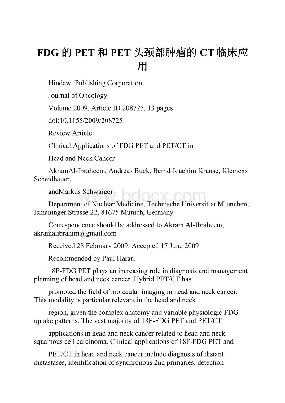
Figure1:
A61-year-oldmanwithnasopharyngealSCCandbilateralcervicallymphnodemetastasesunderwentPET/CTforstaging.Axial
PET,CT,PET/CT,andmaximumintensityprojection(MIP)imagesareshown.PET/CTrevealedfocalFDGuptakeintherightliverlobe
indicatinglivermetastasis(black,whitearrows).PET/CTalsorevealedmultiplefocalFDGuptakesinthelumbarspine,sternum,andribs
indicatingmultiplebonemetastases(redarrows).PET/CTwasvaluablefordetectiondistantmetastases.
ofoccultnodaldiseaseinpreviouslyuntreatedpatientand
wouldnothelpthesurgeoninthemanagementstrategy
ofthepatient,particularlyifthestudyisnegative.They
reportedsensitivityandaspecificityrangingfrom67%to
79%and82%to95%,respectively.Falsenegativefindings
werelikelyrelatedtoeitherthepresenceofmicroscopic
metastasesnotdetectedbyPET/CT,orbytheproximityof
nodalmetastasestotheprimar
 升级会员
升级会员 