业务学习 肾肿瘤Word文件下载.docx
《业务学习 肾肿瘤Word文件下载.docx》由会员分享,可在线阅读,更多相关《业务学习 肾肿瘤Word文件下载.docx(19页珍藏版)》请在冰豆网上搜索。
alsocalledtheexcretoryphase):
Thepyelographicphaseishelpfulforproblemsolvingandtodiagnosepotentialmimicsofcysticrenalmasses.
肾盂期(15分钟后,又称做分泌期):
肾盂期有助于诊断隐匿的肾脏囊性病变。
Thepyelographicphasecandistinguishbetweenhydronephrosis(willshowdenseopacificationinthepyelographicphase)andrenalsinuscysts(willnotopacify).
肾盂期可以鉴别肾盂积水(肾盂期时变得浑浊)和肾窦囊肿(不会变得不透明)。
Refluxnephropathymaycauseadilatedcalyxthatcansimulateacysticrenalmassonthenephrographicphase.Thepyelographicphasewouldshowopacificationofthedilatedcalyx.
反流性的肾病可以导致肾盏的扩大,在肾实质期与肾脏囊性病变很类似。
而在肾盂期扩张的肾盏会变的浑浊。
Thepyelographicphaseisalsousefultodemonstrateacalycealdiverticulumandtoshowthe
relationshipofarenalmasstothecollectingsystemforsurgicalplanning.
肾盂期也可以很好的显示肾盂憩室,也可以显示肾脏占位性病变与肾集合系统的关系,为外科手术提供帮助。
•Optionally,avascularphasecanbeperformedforpresurgicalplanning.
视情况而定,外科手术前需做血管造影检查。
Evaluatingenhancement(CTandMRI)
CT和MRI增强检查的表现
•Thepresenceofenhancementisthemostimportantcharacteristictodistinguishbetweenabenignandmalignantnon-fat-containingrenalmass(alesioncontainingintralesionalfatisalmostalwaysabenignangiomyolipoma,evenifitenhances).
在鉴别非含脂的肾脏占位性病变中(含脂肪的多数为血管平滑肌脂肪瘤,尽管有强化),强化后的表现是非常重要的一个特征。
•OnCT,enhancementisquantifiedastheabsoluteincreaseinHounsfieldunitsonpostcontrast
images,comparedtopre-contrast:
<
(lessthan)10HU,Noenhancement;
10–19HU,Equivocalenhancement.;
≥(greaterthanorequalto)20HU,Enhancement.
增强前后的图像CT值对比:
小于10hu为无强化;
10-19hu为疑似强化;
大于等于20hu为强化。
•OnMRI,enhancementisquantifiedasthepercentincreaseinsignalintensityasmeasuredonpost-contrastimages:
15%:
Noenhancement.15–19%:
Equivocalenhancement.≥20%:
Enhancement.
MRI增强检查,前后对比,小于15%为无强化;
15-19%疑似强化;
大于等于20%为强化。
•Lesionsareconsidered“toosmalltocharacterize”ifthelesiondiameterissmallerthantwicetheslicethickness.Forinstance,using3mmslices,alesionlessthan6mmcannotbeaccuratelycharacterizedbasedonattenuationorenhancement.
如果病灶小于两个层面时,没有特征性的表现。
例如,3毫米层厚时,小于6毫米的病灶基于减弱或者增强时,就不能准确的诊断。
Renalmassbiopsy
肾脏占位性病变的活组织切片检查
•Afterfullimagingworkupiscomplete,thereareseveralwell-acceptedindicationsforpercutaneousrenalmassbiopsy:
所有的影像学检查结束后,有几个被广泛接受的适应症,可以进行肾脏占位性病变的经皮穿刺活检。
Indicationsforrenalmassbiopsy
穿刺活检的适应症
•Todistinguishrenalcellcarcinomafrommetastasisinapatientwithaknownprimary.
鉴别肾细胞性肾癌还是转移性肿瘤。
•Todistinguishbetweenrenalinfectionandcysticneoplasm.
鉴别感染还是囊性的病变。
•Todefinitivelydiagnoseahyperdense,homogeneouslyenhancingmass(afterMRIhasbeen
performed),whichmayrepresentabenignangiomyolipomawithminimalfatversusarenalcell
carcinoma.
最终诊断同肾肿瘤同样强化的高密度病变,代表的有含有很少脂肪的血管平滑肌脂肪瘤与肾细胞肾癌。
•Todefinitivelydiagnoseasuspiciousrenalmassinpatientwithmultiplecomorbiditiesforwhomnephrectomywouldbehighrisk.
在具有高风险的肾脏切除手术并伴有多发并发症的病人,可以最终明确疑似的肾肿瘤性病变。
•Toensurecorrecttissuediagnosispriortorenalmassablation.
在占位性病变切除前明确病理组织诊断。
166
Solidrenalmasses
肾脏实性占位
Renalcellcarcinoma(RCC)
肾细胞性肾癌
Renalcellcarcinoma,stage3A:
Coronal(leftimage)andaxialpost-contrastfat-suppressedT1-weightedMRIshowsaheterogeneouslyenhancingmass(yellowarrows)replacingandexpandingmostoftheleftkidney.Contiguoustothemassthereisexpansionandheterogeneousenhancementoftheleftrenalvein(redarrows),representingtumorthrombusandextensionoftherenalcarcinomaintotherenalvein.
3A期的肾细胞肾癌:
冠状位(左)和轴位T1WI压脂后的增强图像示:
大部分的左侧肾脏被不均匀强化的肾肿瘤(黄箭头)取代,邻近肿块的是扩张和不均匀强化的左肾静脉(红箭头),表示左肾静脉癌栓形成和受累。
•Renalcellcarcinoma(RCC)isarelativelyuncommontumorthatarisesfromtherenaltubularcells.Itrepresents2–3%ofallcancers.RiskfactorsfordevelopmentofRCCincludesmoking,acquiredcystickidneydisease,vonHippel–Lindau(VHL),andtuberoussclerosis.
肾细胞肾癌是起源于肾小管细胞的不是很常见的肿瘤。
在所有肿瘤中占2-3%。
危险因素包括吸烟、继发于肾脏囊性病变、“希佩尔-林道综合征”和结节性硬化。
•ClearcellisthemostcommonRCCsubtype(~75%),withapproximately55%5-yearsurvival.
75%的肾癌为透明细胞癌,其5年存活率接近55%。
ClearcellRCCtendstoenhancemoreavidlythanthelesscommonsubtypes.
透明细胞肾癌相对于其它亚型的肿瘤强化明显。
ClearcellcanbesporadicorassociatedwithvonHippel–Lindau.
透明细胞可以是散发的或者和“希佩尔-林道综合征”相关。
•PapillaryRCCisahypovascularsubtype,witha5-yearsurvivalof80–90%.
乳头状透明细胞癌是少血供的类型,其5年生存率为80-90%。
PapillaryRCCtendstoenhanceonlymildlyduetoitshypovascularity.
乳头状透明细胞癌因为其少血供,表现为轻微强化。
Arenal“adenoma”isfrequentlyseenonautopsyspecimensandisapapillarycarcinoma≤5mm.
肾脏腺瘤通常在尸检标本中发现,死小于5mm的乳头状肾癌。
•Chromophobeisthesubtypewiththebestprognosis,featuringa90%5-yearsurvival.
嫌色细胞癌是一种预后最好的亚型,5年存活率为90%。
•Collectingductcarcinomaisrareandhasapoorprognosis.
集合管癌是少见并预后不良。
•Medullarycarcinomaisalsorare,butisknowntoaffectmostlyyoungadultmaleswithsicklecelltrait.Medullarycarcinomaisanextremelyaggressiveneoplasm,withameansurvivalof15months,nothelpedbychemotherapy.
髓样癌也是少见的,主要发生于具有镰刀型细胞性质的年轻人。
髓样癌是非常有侵袭性的肿瘤,不进行化疗的存活期为15月。
•StagingofrenalcellcarcinomaisbasedontheRobsonsystem,whichcharacterizesfascialextensionandvascular/lymphnodeinvolvement.StagesI–IIIareusuallyresectable,althoughthesurgicalapproachmayneedtobealteredforvenousinvasion(stagesIIIAandIIIC).
肾癌的分级是基于罗布森系统,包括筋膜的受累、血管及淋巴结的转移。
1-3级的通常可以切除,因为静脉的受累,手术入迳常常需要更改。
StageI:
Tumorconfinedtowithintherenalcapsule.
1期:
肿瘤局限于肾包膜内。
StageII:
TumorextendsoutoftherenalcapsulebutremainsconfinedwithinGerota’sfascia.
2期:
肿瘤突破肾包膜,但仍然局限于肾前筋膜。
StageIII:
Vascularand/orlymphnodeinvolvement.
3期:
血管和/或淋巴结转移。
IIIA:
RenalveininvolvementorIVCinvolvement.
IIIA期:
深静脉受累或者下腔静脉受累。
IIIB:
Lymphnodeinvolvement.
IIIB:
淋巴结转移。
IIIC:
Venousandlymphnodeinvolvement.
IIIC:
静脉和淋巴结转移。
StageIVA:
TumorgrowththroughGerota’sfascia;
IVA期:
肿瘤突破肾前筋膜生长。
StageIVB:
Distantmetastasis.
IVB:
远处转移。
167
Angiomyolipoma(AML)
血管平滑肌脂肪瘤
Axialnon-contrastCTshowsanexophyticmass(arrow)intherightkidneycontainingmacroscopicfat.Thereareafewlinearstrandsofsofttissuewithinthelesion.
轴位平扫可见右肾含脂肪的外生性肿块,病灶内含有一些少许软组织密度影。
AxialT1-weightedMRIshowsthatthelesionispredominantlyisointensetointra-abdominalfat.
轴位T1加权MRI示:
病灶为主要表现为同腹腔脂肪相等的信号。
Axialearlyarterialpost-contrastT1-weightedfatsuppressedimageshowsslightenhancementofthe
softtissuecomponents.
动脉增强早期T1脂肪抑制图像示:
软组织成分的轻微强化。
Latearterialpost-contrastT1-weightedfatsuppressedimageshowsmoreprominentenhancementofthesofttissuecomponentsofthelesion.
动脉晚期示:
病变内软组织成分明显强化。
•Angiomyolipoma(AML)isthemostcommonbenignrenalneoplasm,composedoffat,smoothmuscle,anddisorganizedbloodvessels.Themajorityaresporadic,but40%areassociatedwithtuberoussclerosis(whereAMLsarebilateral,withmultiplerenalcysts).
血管平滑肌脂肪瘤是最常见的肾脏良性肿瘤,由脂肪、平滑肌和不规则的血管组成。
大多数是散在的,但是40%和结节性硬化有关(病灶为双侧,伴有多发肾囊肿)。
•AMLhasariskofhemorrhagewhenlarge(≥4cm),thoughttobeduetoaneurysmalchangeofthevascularelements.Small,asymptomaticAMLsarenottypicallyfollowedorresected.
血管平滑肌脂肪瘤大于4cm时有出血的风险,认为是由于血管原因的血管瘤。
小的,无症状的血管平滑肌脂肪瘤通常不需要随访和手术切除。
•Aearlypathognomonicimagingfindingisthepresenceofmacroscopicfatina
non-calcifiedrenallesion.Thenonfat-containingportionenhancesavidlyand
homogeneously.Calcificationisalmostneverpresent.
典型的征象是在无钙化的肾脏病灶内发现脂肪。
不含脂肪的成分明显强化,钙化基本看不见。
•OnMRI,thefatcomponentwillfollowretroperitonealfatonallsequencesandwillsaturateoutonfat-saturatedsequences.IntracytoplasmiclipidisnotafeatureofAML,sothereshouldbenosignificantsignaldrop-outondual-phaseMRI.
磁共振图像,脂肪部分同腹膜后的脂肪一样在stir序列表现为信号降低。
胞质内的脂肪并不是血管平滑肌脂肪瘤的特点,因此在双期磁共振上没有重要的信号减低。
•Approximately4%ofAMLswillnotcontainvisiblemacroscopicfatandwillappearasahyperdenseenhancingmass.MRIisthenextstep,withtheT2-weightedimagesthemostusefultodistinguishfromrenalcellcarcinomainsomecases.
大约4%的血管平滑肌脂肪瘤不含有脂肪,只表现为增强后高信号肿块。
在有些病例可以通过MRI的T2图像来和肾癌鉴别。
AT2hyperintensemasssuggestsRCC(clearcellsubtype)andthepatientcanproceedtosurgery.
T2高信号肿块提示为肾癌(透明细胞),建议病人手术。
AT2hypointensemassisnonspecificandcanrepresenteitherRCC(papillarytype)orAMLwith
minimalfat.AlthoughanAMLtypicallywouldenhancemoreavidlythanapapillaryRCC,biopsyis
warrantedfordefinitivediagnosis.
T2为低信号肿块没有特异性,可以是肾癌(乳头状肾癌)或者血管平滑肌脂肪瘤。
尽管血管平滑肌脂肪瘤比乳头状瘤强化更明显,病例始终是金标准。
•AMLappearshyperechoiconultrasound,althoughupto1/3ofrenalcellcarcinomasmayalsobehyperechoicandultrasoundisthusunreliabletodistinguishAMLfromRCC.
血管平滑肌脂肪瘤在超声上是强回声,1/3的肾癌也是强回声,因此超声用来鉴别肾癌并不可靠。
168
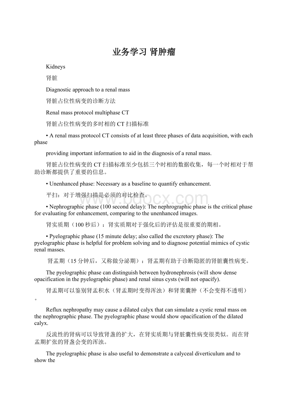
Oncocytoma
嗜酸细胞瘤
Oncocytoma.NoncontrastCT(leftimage)showsanisodenserenalmass(yellowarrows)containingacentralpunctatefocusofhyperattenuation(redarrow).Thecontrast-enhancedpyelographicphaseCT(rightimage)demonstratesthatthemassenhances.Thereisafaintsuggestionofacentralfocusofnonenhancement(redarrow),correspondingtoacentralscar.
嗜酸细胞瘤,平扫CT(左图)表现为等密度肿块(黄色箭头),包含一个中央的点状高密度(红色箭头),增强扫描肾盂期(右图)肿块强化,中心小点状的无强化可以轻微的提示此病,表现为中心瘢痕。
•Oncocyt
 升级会员
升级会员 