分子生物学Chapter 3 Nucleic Acids and Genomics.docx
《分子生物学Chapter 3 Nucleic Acids and Genomics.docx》由会员分享,可在线阅读,更多相关《分子生物学Chapter 3 Nucleic Acids and Genomics.docx(55页珍藏版)》请在冰豆网上搜索。
分子生物学Chapter3NucleicAcidsandGenomics
Chapter3NucleicAcidsandGenomics
Reviewofnucleicacids
ThestructureofDNA,Deoxyribonucleicacid,wasfirstpublishedintheBritishjournalNaturein1953byJ.D.WatsonandF.H.Crick.However,theconstituentsofDNAhadbeenknownsincetheturnofthecentury.ThesimplemodelproposedbyWatsonandCrickimplementedX-raycrystallographyperformedbyM.H.F.WilkinsandR.Franklin.Additionally,workbyL.Paulingprovidedtherulesofbondingandtheelucidationofthealphahelixstructure.E.Chargraffhadprovidedthedataontheabundanceofthefournucleotides(Adenine,Thymine,Guanine,andCytosine)andtherelationshipsbetweenthemfromchemicalanalysis.E.ChargraffdeterminedthatdistributionofAandTwereproportional,andthatdistributionofGandCwerealsoproportionalsuggestingthecomplementaryarrangementofadenine-thymineandguanine-cytosine.SincethearrangementofDNAwascomplementary,thesequenceofonechainmustbecompatibletothesequenceoftheoppositechain.
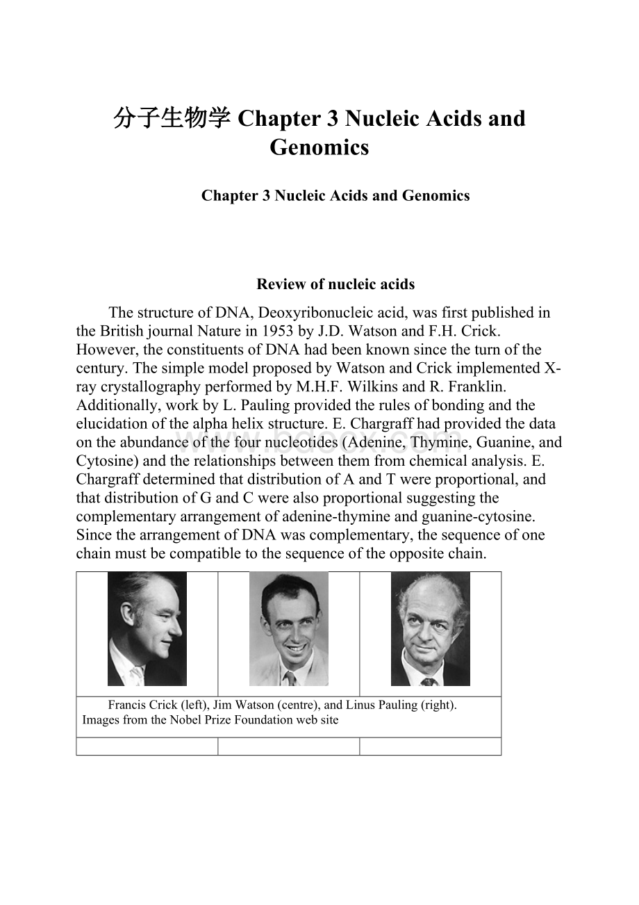
FrancisCrick(left),JimWatson(centre),andLinusPauling(right).
ImagesfromtheNobelPrizeFoundationwebsite
MauriceWilkinsshowedadiffractionpatternofDNAatascientificmeetinginNaplesin1951.ThiswasthefirstdiffractionpatternthatWatsonsawofDNAandithadadecisiveimpactinhisdecisiontostudyDNA.WatsonmovedtotheCavendishlaboratoryinCambridgewherehestruckuphisfamouscollaborationwithFrancisCrick.
Theirfirstattemptatamodelstructure,latein1951,waswrong-embarassinglysoforthematthetime,sincetheyhadarrangedforMauriceWilkinsandRosalindFranklintotravelupfromLondontoviewtheirstructure.Theirmodelwasatriplehelixinwhichthepolynucleotidebackboneswereplacedatthecentreofthestructurewiththebasespointingoutinsolution.But,thisarrangementofchainswaschemicallyimpossibleandoncethiswaspointedout,themodelfellapart.
X-RayfibrediffractionpattersofA-DNA(left)andB-DNA(right).
ImagesfromtheMauriceWilkins1952NobelLectureattheNobelPrizeFoundationwebsite
Chapter3NucleicAcidsandGenomics
1.BuildingBlocks–Nucleotides
vAnucleotideiscomposedofthreeparts:
pentose,baseandphosphategroup.
vA,C,GandT existinDNA;
vA,C,GandU existinRNA.
Bases:
BicyclicPurines:
Monocyclic
pyrimidine:
Nucleosides:
Thebasesarecovalentlyattachedtothe1’positionofapentosesugarring,toformanucleoside
Nucleotides:
Anucleotideisanucleosidewithoneormorephosphategroupsboundcovalentlytothe3’-,5’,or(inribonucleotidesonly)the2’-position.Inthecaseof5’-position,uptothreephosphatesmaybeattached.
BASES
NUCLEOSIDES
NUCLEOTIDES
Adenine(A)
Adenosine
Adenosine5’-triphosphate(ATP)
Deoxyadenosine
Deoxyadenosine5’-triphosphate(dATP)
Guanine(G)
Guanosine
Guanosine5’-triphosphate(GTP)
Deoxy-guanosine
Deoxy-guanosine5’-triphosphate(dGTP)
Cytosine(C)
Cytidine
Cytidine5’-triphosphate(CTP)
Deoxy-cytidine
Deoxy-cytidine5’-triphosphate(dCTP)
Uracil(U)
Uridine
Uridine5’-triphosphate(UTP)
Thymine(T)
Thymidine/
deoxythymidie
Thymidine/deoxythymidie
5’-triphosphate(dTTP)
Phosphodiesterbonds&primarysequence:
Primarysequence:
5’end:
notalwayshasattachedphosphategroups
3’end:
freehydroxyl
(-OH)group
2.DNAStructure
•WatsonandCrick,1953
•Thegeneticmaterialofallorganismsexceptforsomeviruses
•Thefoundationofthemolecularbiology
Basepairing
·TwoseparatestrandsAntiparellel(5’3’direction)
Complementary(sequence)
Basepairing:
hydrogenbondingthatholdstwostrandstogether
•Sugar-phosphatebackbones(negativelycharged):
outside
•Plannerbases(stackoneabovetheother):
inside
2.1DNA'sBForm,AFormandZForm
vInthisstructure,alsoknownastheBform,thehelixmakesaturnevery3.4nm,andthedistancebetweentwoneighboringbasepairsis0.34nm. Hence,thereareabout10pairsperturn. Theintertwinedstrandsmaketwogroovesofdifferentwidths,referredtoasthemajorgrooveandtheminorgroove,whichmayfacilitatebindingwithspecificproteins.
vThenormalright-handed"doublehelix"structureofDNA,alsoknownastheBform
v Inasolutionwithhighersaltconcentrationsorwithalcoholadded,theDNAstructuremaychangetoanAform,whichisstillright-handed,butevery2.3nmmakesaturnandthereare11basepairsperturn.
FeaturesoftheWatson-CrickmodelofB-DNA:
vItisanantiparalleldoublehelix.
vItisaright-handedhelix.
vThebase-pairsareperpendiculartotheaxisofthehelix.(Actually,theyareveryslightlytilted-atanangleof4degrees)
vTheaxisofthehelixpassesthroughthecentreofthebasepairs.
vEachbasepairisrotatedby36degreesfromtheadjacentbasepair.
vThebase-pairsarestacked0.34nmapartfromoneanother.
vThedoublehelixrepeatsevery3.4nm,i.e.thepitchofthedoublehelixis3.4nm.
vB-DNAhastwodistinctgrooves:
aMAJORgroove;and,aMINORgroove.Thesegroovesformasaconsequenceofthefactthatthebeta-glycosidicbondsofthetwobasesineachbasepairareattachedonthesameedge.However,becausetheaxisofthehelixpassesthroughthecentreofthebasepairs,bothgroovesaresimilarindepth.
TherealstructureofB-DNA
WatsonandCrick'sstructurewasajustamodel-butaprettygoodone.Nevertheless,ittooknearly30yearsbeforethestructuresofDNAwereresolvedatatomicresolution.
In1980,RichardDickersonandHoraceDrewsolvedthestructureofa12-merdouble-strandedself-complementaryoligonucleotidewiththefollowingsequence:
5'-CGCGAATTCGCG-3'
TheirresultsshowedthatcrystalsofB-DNAhadastructureverysimilartothatproposedbyWatsonandCrick.Althoughtherewerenumeroussmallvariations,theoverallstructurewasasexpected.
∙Themoleculewasaright-handeddouble-helix.
∙Thebackbonechainswereantiparallel.
∙Thebasepairswereverynearlyperpendiculartothehelixaxis.
∙Thebasepairswerecentredonthehelixaxis.
∙Onaverageeachbasepairwasrotated35.6degreesfromtheadjacentbasepair.However,theindividualmeasuredrotations(twist)variedfromaslittleas28degreestoasmuchas42degrees.
Onaveragetheriseperbasepair(i.e.thedistancebetweenadjacentbasepairs)was0.34nm.However,therisebetweenindividualbasepairsvariedfrom0.274nmto0.435nm.
OneofthemoststrikingfeaturesofthestructuresolvedbyDickersonandDrewwasthatthereisconsiderablevariationinthestructureofindividualbasepairs.Manywerewerenotexactlyplanarbutwereslightlytwisted(propellortwist).Thisfeature,aswellasthevariationsinthetwistanglesandtherisebetweenbasepairscanbeexplainedorunderstoodasresultingfromthecomplexinterplayofattractiveandrepulsiveforcesduetothedifferentchemicalpropertiesofthefourdistinctbaseswithinaDNAdoublehelix.ThedetailedstructureofaDNAmoleculeisactuallyinfluencedbythesequence.
A-DNA
RecallthatthefirstfibrediffractionpicturestakenbyRosalindFranklinwereofadehydratedformofDNA,whichwenowknowasA-DNA.
AneasywaytorememberthebasicstructureofA-DNAistorememberthisfactaboutitswatercontent.InA-DNAthereisnowaterspine.Thebasepairs--whichneverthelessareformedwithcanonicalWatson-Crickhydrogenbonds--arepushedtowardstheminorgroove.Indoingso,thebasepairstiltto19degreesfromperpendiculartothehelixaxiswhichnolongerpassesthroughthecentreofeachbasepair.Theresultingminorgrooveisaboutaswideastheresultingmajorgroove.However,themajorgrooveisverydeepwhiletheminorgrooveisveryshallow.Inaddition,thesugarringchangesfromtheC2'-endoconformationfoundinB-DNAtoaC3'-endoconformation.ThischangeservestodistancethephosphatefromtheC2'hydroxylatomhencepreventingautocatalysis
ThefollowingimagesshowthestructureofA-DNA.
Notethefollowingabouttheseimages:
∙ThefirstimageshowstheA-DNAhelixfromtheside.Noticethatthebasepairsaretiltedwithrespecttotheaxisofthehelix.Mostofthebasepairsarenearlyplanar,but,aswithB-DNA,youcanseesomeexceptions.Youcanalsoseesomeindicationofpropellortwist.
∙Observethebasepairthatishalfwayalongthehelix.Yourviewofthisbasepairisessentiallyend-on.Fromthisperspective,youcanseethatthisbasepairisnotcentredonthehelixaxisbutisverymuchpushedtooneside.Themajorgroove(ontheright)isverydeep;theminorgroove(ontheleft)isveryshallow.
∙ThesecondimageshowstheA-DNAhelixfromoneend.Noticethatthereisessentiallyalarge"hole"inthecentrebecausealloftheatomsofthebasepairsarepushedouttothesides.
AnotherDNAstructureiscalledtheZform,becauseitsbasesseemtozigzag. ZDNAisleft-handed. Oneturnspans4.6nm,comprising12basepairs.
vTheDNAmoleculewithalternatingG-Csequencesinalcoholorhighsaltsolutiontendstohavesuchstructure.
Z-DNA
EversinceWatsonandCrickproposedtheirstructureforDNA,crystallographicproofwassought.In1979,AlexRichandhiscolleaguesatMITthoughttheyhadit!
Theysucceededincrystallizingtheself-complementaryhexanucleotide,CGCGCG.Theiranalysiswasasurprise.
Theircrystalsshowedthatthisparticularmoleculeadoptedaleft-handeddoublehelixundertheircrystallizationconditions.Thebaseshadadoptedasyn-conformationratherthantheusualanti-conformationwiththeresultthattherepeatingunitofthestructurewasadinucleotidebase
 升级会员
升级会员 