组胚切片报告.docx
《组胚切片报告.docx》由会员分享,可在线阅读,更多相关《组胚切片报告.docx(23页珍藏版)》请在冰豆网上搜索。
组胚切片报告
组织学与胚胎学
实验报告
指定主题:
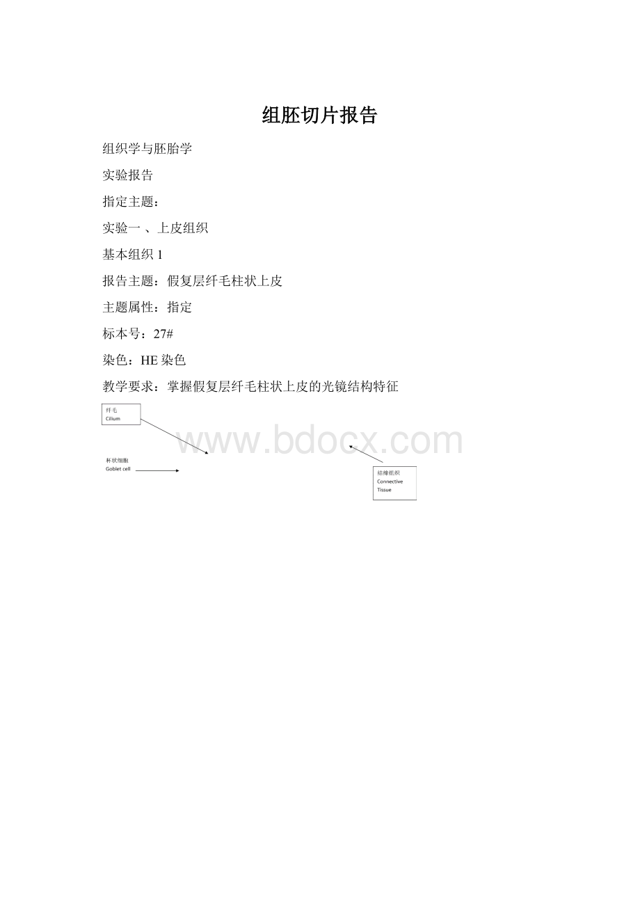
实验一、上皮组织
基本组织1
报告主题:
假复层纤毛柱状上皮
主题属性:
指定
标本号:
27#
染色:
HE染色
教学要求:
掌握假复层纤毛柱状上皮的光镜结构特征
光镜下可见假复层纤毛柱状上皮的各种上皮细胞界限不清,细胞核染蓝紫色,高矮不一,其中柱状细胞最多,游离面有大量纤毛,柱状细胞之前常夹有杯状细胞,其底部狭窄,顶部膨大,细胞核深染。
所有细胞的基底膜均附着在基膜上,即使细胞核高低不齐排列,形似复层,实为单层。
Thenvisiblepseudostratifiedciliatedcolumnarepitheliumofepithelialcelllineisnotclear,thenucleusdyedpurpleblue,heightisdiffer,thecolumnarcellsmost,freesurfacehasalotofciliatedcolumnarcells,beforeoftencliphasgobletcell,itsbottomisnarrow,thetopswollen,withhyperchromaticnuclei.Allthecellsofthebasementmembraneareattachedonthebasementmembrane,whosenucleusareirregular,andtheshapeseeascompoundlayer,anditisasingle.
实验二结缔组织
基本组织2
报告主题:
骨单位
主题属性:
指定
标本号:
6#
材料:
骨切片
染色:
Schmorl氏法块染
教学要求:
掌握骨组织的结构
图2骨单位(骨切片,Schmorl氏法块染,40×100)
Figure2osteon(osseoussmear,Schmorlstained,40×100)
骨单位位于内外环骨板之间,数量最多,中央为圆心的中央管,中央管与穿通管相通,数层同心圆排列的骨板围绕中央管。
在骨单位之间,有一些不规则排列的间骨板,其中无血管通道,在骨板之间或骨板中可见黄色小腔,即骨陷窝。
Osteonislocatedintheinsideandoutsideringboneplatebetween,thelargestnumber,thecentraltotherightofthecentraltube,thecentraltubeandpiercedtubeareinterlinked,layersofconcentricarrangementoftheboneplatearoundthecentralcanal.Therearesomeinterstitiallamellaebetweenosteon,whichhasnovascularchannels,intheboneplateorbetweenboneplatevisibleyellowsmallcavities,namedlacuna.
实验三、肌组织与神经组织实验二
基本组织3
报告主题:
心肌
主题属性:
指定
标本号:
12#
材料:
人心肌切片
染色:
HE染色
教学要求:
观察人心肌的结构特点,掌握其判断依据
图3心肌(人心肌切片,HE染色,40×100)
Figure3Cardiacmuscle(humancardiacmusclesmear,HEstained40×100)
多数心肌纤维有一个细胞核,少数有双核,核呈卵圆形,位于细胞中央。
心肌纤维的连接处称闰盘,且染色深。
心肌纤维呈明暗相间的周期性横纹,但由于其肌原纤维粗细不等,界限不明,因此横纹不如平滑肌那么清楚。
Mostofthemyocardialfibershaveanucleus,afewhavedual-core,andnucleusisoval,locatedinthecentralcell.Thejointofthemyocardialfiberscalledintercalateddisc,anddeepdyeing.Themyocardialfibersarelightandshadeandperiodichorizontallines,butasaresultofitsmyofibrildifferentthicknessesoftheunknown,sohorizontalstripesassmoothmusclesoclear.
基本组织4
报告主题:
多级神经元
主题属性:
指定
标本号:
9#
材料:
小牛脊髓及神经节
染色:
HE染色
教学要求:
观察脊髓前角多级神经元的结构特点,掌握其判断依据
图4多级神经元(小牛脊髓切片,HE染色,40×100)
Figure4multipolarneuron(calfspinalcordsmear,HEstained40×100)
多级神经元细胞核有大、圆,亮的特点,胞体和突起中可见到许多紫蓝色的不规则的小斑块,即尼氏体。
突起从胞体发出,一个神经元有多个树突,一个轴突,因此轴突很难切到,不容易看见。
Multistageneuronsnucleihavebig,round,brightcharacteristics,cellandbumpsinthevisibletomanypurpleblueirregularsmallpatches,namelytheNisslbody.Flashfromthecellbodyout,aneuronhavemultipledendrite,anaxon,soitisdifficulttocuttheaxon,whichisnoteasytosee.
实验四消化管
中空性器官1
报告主题:
空肠绒毛
主题属性:
指定
标本号:
19#
材料:
猫空肠横切片
染色:
HE染色
教学要求:
掌握空肠的结构特征与判断依据,特别注意肠绒毛和肠腺的结构)
图5空肠绒毛(猫空肠横切片,HE染色,40×100)
Figure5jejunumvilli(catjejunumtransversesection,HEstained40×100)
空肠绒毛上皮由吸收细胞、杯状细胞和少量内分泌细胞组成。
吸收细胞数量最多,呈高柱状,核长椭圆型,位于基部,细胞游离有深染层,称纹状缘。
绒毛中轴的固有层为细密结缔组织,其内常可见纵行的平滑肌、丰富毛细血管、1~2条中央乳糜管(多数未切到)及大量淋巴细胞。
TheepitheliumofJejunumvilliiscomposedofabsorption,gobletcellandasmallamountofendocrinecells.Thelargestnumberofabsorptivecells,atallcolumnar,nuclearlongelliptic,islocatedinthebase,cellfreehavehyperchromaticlayer,calledstriatedborder.Villiaxisofthelaminapropriaforfineconnectivetissue,itoftenvisiblelongitudinaldonesmoothmuscle,richcapillaries,1~2acentrallacteal(mostnotcut)andalargenumberoflymphocytes.
实验五消化管以外的其他中空性器官
中空性器官2
报告主题:
中等动脉
主题属性:
指定
标本号:
10#
材料:
狗中等动、静脉横切片
染色:
HE染色
教学要求:
掌握中等动脉的结构特点
图6中等动脉(狗中等动脉横切片,HE染色,40×100)
Figure6medium-sizedartery(dogmedium-sizedarteryandveintransversesectionsmear,HEstained40×100)
在高倍镜下,可见内弹性膜为一条呈波浪状的折光性强的粉红色亮带,它是内膜与中膜的分界标志,中膜较厚,由数十层平滑肌纤维组成,因此中动脉也称肌性动脉。
外膜由疏松结缔组织构成,多数中动脉的外膜和中膜之间有明显的外弹性膜。
Athighmagnification,internalelasticmembraneisawavyrefractionsexstrongpinklightband,whichistheborderoftunicamediaandtunicaadventitia.tunicamediaisconsistofdozensoflayerofthesmoothmusclefibers,sothearteryisalsocalledthemuscularartery.Externalelasticmembraneiscomposedoflooseconnectivetissue,themajorityoftheartery;thereisobviouselasticmembranebetweentunicamediaandtunicaadventitia.
实验六免疫系统
实质性器官1
报告主题:
淋巴结
主题属性:
指定
标本号:
13#
材料:
狗淋巴结切片
染色:
HE染色
教学要求:
掌握淋巴结的结构特征和判断依据
图7淋巴结(狗淋巴结切片,HE染色,40×100)
Figure7Lymphoidnode(Doglymphoidsmear,HEstained,40×100)
淋巴结多呈扁平豆形或者卵圆形,表面有薄层结缔组织被膜,伸入实质形成小梁,小梁互相连接成网。
皮质位于被膜下,浅皮质由淋巴小结和弥散淋巴组织构成,副皮质为胸腺依赖区,为较大片的弥散淋巴组织,皮质淋巴窦位于被膜下方和小梁周围.髓质由髓索及其间的髓窦构成。
Lymphnodeisflatbeanshapedorovoid,thesurfacehasathinlayerofconnectivetissuecapsule,stretchintosubstantialformtrabecular,trabecularinterconnectedintonetwork.Cortexislocatedintheenvelope,shallowcortexbylymphnoduleanddispersionlymphoidtissuestructure;vicecortexforthymusdependonthearea,foralargedispersionlymphoidtissue,cortexlymphaticsinus,intheenvelopebelowandtrabecularmeshwork.Medullaaroundbypulplineandthepulpsinusbetweencompositions.
实验八肾与内分泌系统
实质性器官2
报告主题:
下颌下腺
主题属性:
自选
标本号:
24#
材料:
人下颌下腺切片
染色:
HE染色
教学要求:
掌握下颌下腺的三种腺泡和导管的结构
图8下颌下腺(人下颌下腺切片,HE染色,40×100)
Figure8(Submandibularglandsmear,humansubmandibulargland,HEstained40×100)
下颌下腺为混合性腺,小叶内可见很多圆形或不规则的腺泡,其中染紫红色的是浆液性腺泡,染色浅的是黏液性腺泡,混合性腺泡主要由黏液性腺细胞构成,少量浆液性腺细胞位于黏液性细胞之间或聚集在腺泡底部,也称为半月。
分泌管为单层柱状上皮,多且发达,闰管短而不明显。
Thesubmandibularglandismixedgland,theleavescanbeseenmanyroundorirregularacinus,includingdyeaborigineisserousacinus,dyeingshallowismucousacinus,mixedglandismainlycomposedofmucouscellsandasmallamountofserouscellsintheslimesexbetweencellsorgatheredinglandbubblebottom,calleddemilune.Secretaryductforsinglecolumnarepithelium,anddeveloped,leaptubeshortisnotobvious.
实验八肾与内分泌系统
实质性器官3
报告主题:
肾单位
主题属性:
指定
标本号:
29#
材料:
兔肾切片
染色:
HE染色
教学要求:
掌握肾单位各部分的结构特点
图9肾单位(兔肾切片,HE染色,40×100)
Figure9Nephron(Rabbitkidneysmear,HEstained,40×100)
肾单位由肾小体和与其相连的肾小管组成,肾小体由血管球和肾小囊构成,血管球为肾小囊内一团蟠曲的毛细血管,肾小囊是上皮性管道盲端膨大并凹陷的双层囊。
近曲小管位于肾小体周围,管径大,管壁厚,管腔小而且不规则。
远曲小管与近曲小管混在一起,管径小,管壁薄,管腔较大而规则。
Nephroniscomposedofrenalcorpuscleandrenaltubule.Renalcorpusclebyvascularballandrenalcapsuleform,glomusforrenalcapsuleinagroupofcapillary,renalcapsuleisdoublecapsule.Proximalconvolutedtubuleinrenalcorpusclearound,pipediameter,wallthickness,thelumenissmallandirregular.Distalconvolutedtubuleandproximalconvolutedtubulemixedtogether,pipediameterissmall,andthewallisthin
实验九生殖腺
实质性器官4
报告主题:
生精小管
主题属性:
指定
标本号:
41#
材料:
人睾丸与附睾切片
染色:
HE染色
教学要求:
了解睾丸与附睾的一般结构,掌握生精小管的结构和间质细胞的位置及其结构特点。
图10生精小管(人睾丸与附睾切片,HE染色,40×100)
Figure10Somniferoustubule(humantestisandepididymis,HEstained,40×100)
生精小管为高度弯曲的复层上皮性管道,外有薄层基膜,管壁由各级生精细胞和支持细胞构成。
精原细胞附着在基膜上,胞体较小,核染色较深。
初级精母细胞位于精原细胞近腔侧,细胞体积大而圆,核亦大而圆,染色质密集呈绒球状。
次级精母细胞因存在时间短不易找到,精子细胞位于近腔面,体积更小,核大而圆,染色质细密。
精子位于腔面,头部呈扁圆形,嵌入支持细胞的胞质中,染色深,尾部游离于生精小管腔内。
Somniferoustubulesarecurvedstratifiedepitheliumofpipeline,outsideathinlayerofbasementmembrane,wallbyvariousrawspermcellandsupportcellscomposition.Spermatophoreattachedonthebasementmembrane,cellislesser,nuclearstainingdeep.Primaryspermatocytelocatedinnearlycavityside,cellvolumeisbigandround,nuclearalsolarge,round,chromatinconcentratedinwoolball.Secondaryspermatocytethereforshorttimeisnoteasytofind,spermcellsinthenearlycavitysurface,thevolumeissmaller,nuclearlarge,round,chromatinclose.Spermislocatedinthecavitysurface,theheadisoblateness,embeddedsupportcellcytoplasm,dyeingdeep,tailfreeborninsomniferoustubulecavity.
自选主题
实验二、结缔组织
基本组织5
报告主题:
嗜酸性粒细胞
主题属性:
自选
标本号:
7#
材料:
人血涂片
染色:
Wright染色
教学要求:
掌握嗜酸性粒细胞
图11嗜酸性粒细胞(人血涂片,HE染色,40×100)
Figure11Esoinophil(humanbloodsmear,HEstained,40×100)
嗜酸性粒细胞核多分为两叶,胞质内充满粗大、分布均匀、染成橘红色、有折光性的嗜酸性颗粒。
其主要作用是能吞噬抗原抗体复合物,分解组胺,灭活白三烯,从而减弱过敏反应,又参与对蠕虫的免疫反应。
Eosinophilicnucleararemainlydividedintotwoleaves,cytoplasmfilledwithbulky,distributionuniformity,dyedorange,refractionsexeosinophilicparticles.Itsmainfunctionistodevourantigenantibodycomplex,decompositionofhistamine,inactivatedleukotriene,therebydeadeningallergicreaction,andthewormintheimmuneresponse.
实验一、上皮组织
基本组织6
报告主题:
变移上皮
主题属性:
自选
标本号:
31#
染色:
HE染色
材料:
猫膀胱切片(空虚状态)
教学要求:
掌握膀胱空虚状态下变移上皮的形态结构
图12变移上皮(猫膀胱切片,HE染色,40×100)
Figure12Transitionalepithelium(catbladdersmear,HEstained,40×100)
变移上皮由多层细胞构成,基底面平整。
细胞核常排成5~6层,其中,基底层细胞核密集,小且染色深;中间数层细胞多为多边形或倒梨形,核圆;表层细胞大,分界清晰,有防止尿液侵蚀的作用,称为盖细胞。
Transitionalepitheliumcellsbymultilayerstructure,basewithsmoothsurface.Thenucleusisoftenin5~6layer,amongthem,thebasallaminanucleiintensive,smallanddyeingdeep;Intermediatelayersofcellsforpolygonorfallpear-shaped,nuclearround;Thesurfacecells,boundaryclear,havepreventurineerosioneffect,calledcovercell.
实验四、消化管
中空性器官3
报告主题:
食管
主题属性:
自选
标本号:
18#
染色:
HE染色
材料:
狗食管和胃纵切片
教学要求:
掌握食管的结构特点及判断依据
图13食管(狗食管和胃纵切片,HE染色,40×100)
Figure13Esophagus(dogesophagusandstomachlongitudinalsection,HEstained,40×100)
食管黏膜的上皮为复层扁平上皮,固有层为细密结缔组织,黏膜肌层由纵行平滑肌束组成。
黏膜下层的结缔组织中存在许多黏液性的食管腺。
肌层由内环形和外纵行两层,人的食管上段为骨骼肌,下段为平滑肌。
外膜为纤维膜。
Esophagealmucosaepithelialisstratifiedsquamousepithelium,Muscularismucosaisconnectivetissue,longitudinalbeamofsmoothmusclemakeupmuscularismucosa.Submucosaofconnectivetissuehasmanyesophagealglands.musularisfrominnerringandouterlongitudinaltwolayer,humanesophagealuppersegmentforskeletalmuscle,lowersegmentforsmoothmuscle.Tunicaadventitiaisfibermembrane.
实验七、消化腺与肺
实质性器官5
报告主题:
肝
主题属性:
自选
标本号:
26#
染色:
HE染色
材料:
香猪肝切片
教学要求:
掌握肝的结构特点及其判断依据。
图14肝(香猪肝切片,HE染色,40×100)
Fi
 升级会员
升级会员 