距骨骨折手术入路前外侧入路.docx
《距骨骨折手术入路前外侧入路.docx》由会员分享,可在线阅读,更多相关《距骨骨折手术入路前外侧入路.docx(10页珍藏版)》请在冰豆网上搜索。

距骨骨折手术入路前外侧入路
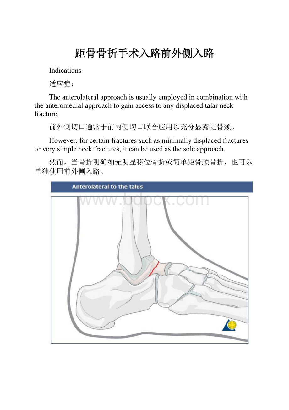
Indications
适应症:
Theanterolateralapproachisusuallyemployedincombinationwiththeanteromedialapproachtogainaccesstoanydisplacedtalarneckfracture.
前外侧切口通常于前内侧切口联合应用以充分显露距骨颈。
However,forcertainfracturessuchasminimallydisplacedfracturesorverysimpleneckfractures,itcanbeusedasthesoleapproach.
然而,当骨折明确如无明显移位骨折或简单距骨颈骨折,也可以单独使用前外侧入路。
Anatomy 解剖
Aroundtheankleandhindfoot,full-thicknessincisionswithoutunderminingareimperative.Toavoidcuttingthebranchesofthesuperficialperonealnerve,mostoftheincisionsmustbemadeinalongitudinaldirection.
在踝关节和后足,必须用全厚层皮瓣切口。
为避免切断腓浅神经,切口需要设计纵轴方向。
Thesuperficialperonealnerveliesintheincisionorverynearandmustbeavoidedandprotected.
腓浅神经多位于手术切口附件,必须注意保护。
Incision 切口
Skinincision 皮肤切口
Theincisionsherearebasedonthedeeptalaranatomywhichliesunderneaththeextensordigitorumbrevis.
Theincisionisbasedonthefourthmetatarsalandlinesupwiththisbone.
切口与其下的解剖相关,伸趾短肌位于下方,与第4跖骨平行。
Deepdissection 深层分离
Themuscleofextensordigitorumbrevisisbulkybutoncethismuscleissplit, onegainsaccesstothelateraltalusandsubtalarjoint.
伸趾短肌体积明显,但是,一旦分离此肌肉,距骨外侧和距下关节就看到距骨前外侧和距下关节了
Exposureoftheanterolateraltalarneck
显露前外侧距骨颈
Oncetheextensordigitorumbrevisissplitlongitudinallyandretracted,oneexposesthelateralaspectofthetalarneck。
当伸趾短肌被牵开,距骨颈就显露出来了。
Mosttalarneckfracturesresultfromamediallydirectedforce.Therefore,thelateralsideoftheneckcomesundertensionandthemedialsideundercompression.Thefracturesonthelateralsidearesimple,andonthemedialsidemultifragmentary.Exposurefromthemedialsidealonerunstheriskofshorteningtheneckandmalreducingthefracture.Toensureananatomicreduction,fracturesoftheneckmustbeexposedfromthelateralandmedialside.
多数距骨颈骨折是由于内侧直接暴力引起。
因此,距骨颈外侧面承受张力,而内侧面承受压力。
所有,外侧面骨折多是简单骨折,内侧面骨折多粉碎骨折。
单纯显露内侧面有引起距骨颈短缩,和引起骨折复位不良。
为了达到解剖复位,颈部骨折必须暴露外侧和内侧面。
Thearrowontheimageindicatesasimplefractureofthetalarneck.
下图的箭头显示距骨颈简单骨折
Debridementofsubtalarjoint 复位距下关节
Debridementofthesubtalarjointisimperativetofacilitateanexcellentreduction.
距下关节必须得到很好的复位。
Usingbothanterolateralandanteromedialapproachesprovidesexcellentaccesstothewholetalarneck.Theimportanceofthisliesingettingaperfectreductionfordefinitivefixation.
同时使用前外和前内切口可以提供足够距骨颈的显露,并可以在很好的复位和固定。
OurCTimageshowscomminutionoftheinferiortalus.
CT显示距下关节粉碎骨折